
Fig. 8
- ID
- ZDB-FIG-181114-8
- Publication
- Sun et al., 2018 - Maternal Ybx1 safeguards zebrafish oocyte maturation and maternal-to-zygotic transition by repressing global translation
- Other Figures
- All Figure Page
- Back to All Figure Page
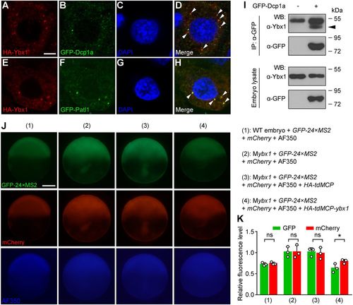
Ybx1 is localized in P-body-like granules in embryos and represses the translation of bound mRNA. (A-H) Colocalization of HA-Ybx1 with the GFP-labelled P-body components Dcp1a (A-D) and Patl1 (E-H) in 4 hpf embryos. HA immunofluorescence (A,E), GFP immunofluorescence (B,F), DAPI staining (C,G) and merged signals (D,H) are shown in confocal slices. Arrowheads indicate colocalization foci. Scale bar: 5 μm. (I) Association of endogenous Ybx1 with GFP-Dcp1a in embryos revealed by co-IP. Embryos were injected with gfp-dcp1a mRNA at the one-cell stage and harvested at 8 hpf. IP, immunoprecipitation; WB, western blot. The arrowhead indicates the band of immunoprecipitated endogenous Ybx1 protein. (J) Fluorescent images showing the levels of GFP-24×MS2, mCherry and AF350 dye (loading control). Embryo genotypes and injected reagents are listed on the right. Scale bar: 200 μm. (K) Measurement of GFP and mCherry fluorescence levels relative to AF350. Both GFP and mCherry levels are upregulated in 2 and 3 compared with 1. Note the marked decrease of GFP intensity and the lesser decrease of mCherry in 4 compared with 3. ns, not significant; *P<0.05; n=3 embryos; Student's t-test. |