
Fig. 7
- ID
- ZDB-FIG-180914-10
- Publication
- Matejčić et al., 2018 - A non-cell-autonomous actin redistribution enables isotropic retinal growth
- Other Figures
- All Figure Page
- Back to All Figure Page
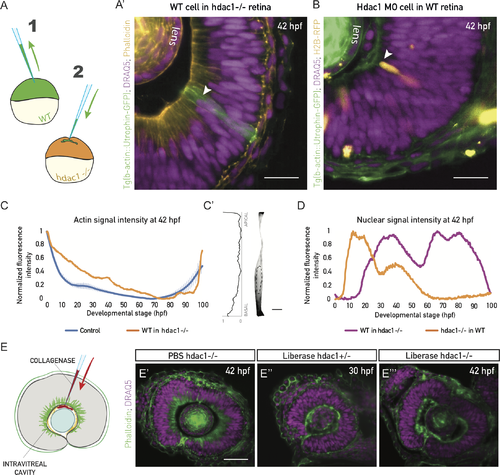
Actin reorganization and cell height increase are non-cell autonomous and ECM dependent. (A) Schematic of the cell transplantation experiment. Blastomeres from Tg(actb1::GFP-UtrCH) embryos (1) were transplanted into acceptor hdac1−/− embryos (2) between high and sphere stage. (A’) WT cells in hdac1−/− tissues do not redistribute basolateral actin, nor do they increase their height at 42 hpf (green cells, arrowhead). Actin is labeled with phalloidin (orange) and nuclei with DRAQ5 (magenta). N = 5 embryos. Scale bar: 50 μm. (B) Mosaic injection of hdac1 MO into Tg(actb1::GFP-UtrCH). Cells with hdac1 MO were co-injected with H2B-RFP mRNA. Their basal actin zone disappears at the same time as in the WT (orange cells, arrowhead). Actin is labeled with phalloidin (green) and nuclei with DRAQ5 (magenta). N = 5 embryos. Scale bar: 50 μm. (C) Normalized fluorescence intensity profiles of phalloidin signal along the apicobasal axis of the transplanted clone marked in (A’). Control plot is same data as Fig 4E. (C’) Phalloidin signal in a manually segmented transplanted PSE cell from sample in (A’), showing basolateral accumulation on the single-cell level. Dashed line outlines the cell nucleus. Scale bar: 5 μm. Left: Normalized basal-to-apical phalloidin signal intensity along the lateral cell membrane of cell on right. (D) Normalized fluorescence intensity profile of nuclear signal (H2B, orange line) along the apicobasal axis of the hdac1-deficient clone in (B) and nuclear signal (DRAQ5, magenta line) in the transplanted clone in (A’). (E) ECM integrity was perturbed by injecting 0.5 mg/ml of collagenase (Liberase) into the intravitreal cavity of the zebrafish eye. (E’) The basal actin accumulation in hdac1−/− tissues remains intact when PBS is injected as control. (E”) Collagenase abolishes the basal accumulation in 30 hpf control hdac1+/− embryos. (E”’) Collagenase abolishes the basal actin accumulation in 42 hpf hdac1−/− tissues. E’-E”’, actin is labeled with phalloidin (green), nuclei are labeled with DRAQ5 (magenta). (Underlying data for panels C and D can be found at DOI: 10.5281/zenodo.1316912; /Matejcic-et-al_2018/Data/F7CD.csv.). DRAQ5, deep red anthraquinone 5; ECM, extracellular matrix; hdac1, histone-deacetylase 1; hpf, hours post fertilization; MO, morpholino; PSE, pseudostratified epithelium; WT, wild type. |