
Fig. 4
- ID
- ZDB-FIG-180717-15
- Publication
- Hendee et al., 2018 - PITX2 deficiency and associated human disease: insights from the zebrafish model
- Other Figures
- All Figure Page
- Back to All Figure Page
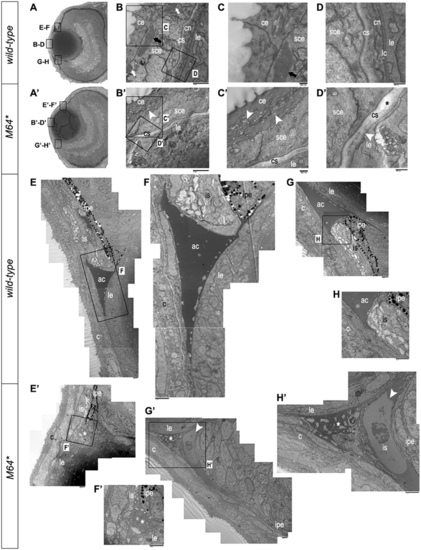
Electron microscopy of 3-dpf anterior chamber structures. (A, A’) Histological sections depicting orientation of wild-type (A) and pitx2M64* mutant (A’) eyes. (B–D, B’–D’) Wild-type (B–D) and pitx2M64* mutant (B’–D’) central corneas. Wild-type eyes display three layers of stratification: epithelium, stroma and endothelium (B). The corneal epithelium is delineated into surface and subepithelial cells distinguished by many dark-staining small inclusion bodies versus fewer larger inclusion bodies, respectively (arrows in B, C). The wild-type corneal stroma is of uniform thickness and on its basal side borders the corneal endothelium (D). In contrast, corneal defects in the mutants include an epithelium that contains smaller inclusion bodies in both layers and exhibits small vacuoles in the surface layer (arrowheads in B’, C’), a thicker and uneven stroma (asterisk in D’), and lack of a defined endothelial layer (arrowhead in D’). (E–H’) Wild-type (E–H) and pitx2M64* mutant (E’–H’) iridocorneal angles. Dorsal (E–F) and ventral (G–H) angles displayed early formation of the anterior chamber space as well as early differentiation of the iris stroma and iris pigmented epithelium. In mutants, both dorsal (E’, F’) and ventral (arrowheads in G’, H’) iridocorneal angles display notable under-differentiation of the iris stroma and iris pigmented epithelium, particularly ventrally; in addition, the anterior chamber space is marked by an excess of cellular material (asterisks in E’, H’). ce, corneal epithelium; cn, corneal endothelium; cs, corneal stroma; lc, lens capsule; le, lens epithelium; sce, corneal subepithelium; ac, anterior chamber; c, cornea; is, iris stroma; ipe, iris pigmented epithelium. |
| Fish: | |
|---|---|
| Observed In: | |
| Stage Range: | Protruding-mouth to Adult |