
Fig. 5
- ID
- ZDB-FIG-180717-16
- Publication
- Hendee et al., 2018 - PITX2 deficiency and associated human disease: insights from the zebrafish model
- Other Figures
- All Figure Page
- Back to All Figure Page
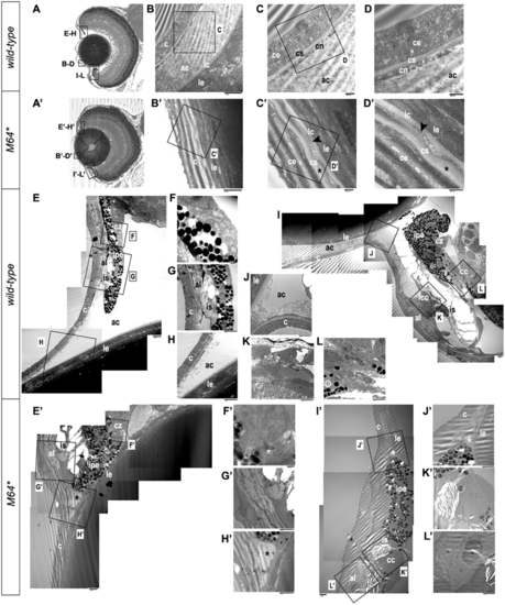
Electron microscopy of 14-dpf anterior chamber structures. Histological sections depicting orientation of wild-type (A) and pitx2M64* mutant (A’) eyes. Wild-type (B–D) and pitx2M64* mutant (B’–D’) central corneas. The wild-type cornea is clearly stratified into its three component layers: a three- to four-cell layer thick epithelium; a smooth, even-thickness stroma; and a uniform single layer endothelium (B–D). The corneal endothelium is distinctly separated from the lens capsule and epithelium by the anterior chamber space (B). In mutants, the anterior chamber space is absent, placing the mutant cornea directly up against the lens capsule (B’–D’). The innermost layer of subepithelium and stroma appear wavy and of fluctuating thickness (asterisks in C’, D’). The presumptive corneal endothelium, be it a monolayer endothelium or the compressed remnants of the anterior chamber space, is contoured between the stroma and lens capsule (arrowheads in C’, D’). Wild-type (E–L) and pitx2M64* mutant (E’–L’) iridocorneal angles. Wild-type dorsal (E–H) and ventral (I–L) angles display a distinct anterior chamber space with separation between lens and cornea and no angle occlusion (E, H–J); in addition, further differentiation of the iris stroma and the presumptive annular ligament is also demonstrated (E, G, I, K). Dorsally, the ciliary zone non-pigmented epithelial cells have begun to anteriorly differentiate from the retinal neuroepithelium (E, F). Ventrally, the formation of the iridocorneal canal and the ciliary canal, the two main branches of the specialized canalicular drainage network, can be observed (I, K, L). In contrast, the mutant shows a notable reduction in the anterior chamber space at both iridocorneal angles, with the iris pigmented epithelium remaining markedly in contact with the lens (asterisks in E’, H’–J’). The iris stroma and presumptive annular ligament appear delayed in their differentiation (E’, G’, I’, K’, L’). Dorsally, the non-pigmented ciliary epithelium does not appear to have begun differentiation (E’, F’). Ventrally, the only indication of specialization is the break in the iris pigmented epithelium marking the presumptive location of the ciliary canal (I’, K’); aside from this, the endothelial lining of the canal has failed to differentiate, and the iridocorneal drainage canal is undetectable (I’, K’, L’). ac, anterior chamber; c, cornea; ce, corneal epithelium; cn, corneal endothelium; cs, corneal stroma; lc, lens capsule; le, lens epithelium; al, annular ligament; cc, ciliary canal; cz, ciliary zone; icc, iridocorneal canal; ipe, iris pigmented epithelium; is, iris stroma. |
| Fish: | |
|---|---|
| Observed In: | |
| Stage: | Days 14-20 |