
Fig. S2
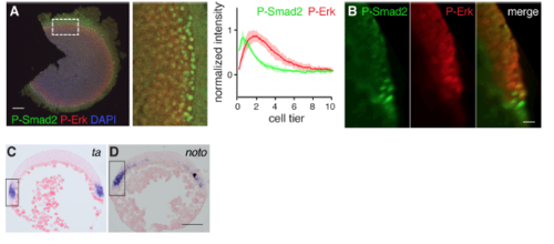
Cells directly adjacent to the YSL maintain low levels of P-Erk (A) Example of flat-mounted, 50% epiboly embryo, stained by immunohistochemistry for PErk and P-Smad2. Right panel shows average of intensity traces of three embryos (mean ± SD). Scale bar, 100 μm. (B) Resliced optical section of 50% epiboly embryo stained as in (A) obtained with light sheet microscope. Note that nuclei close to the YSL have high P-Smad2, but low P-Erk levels. Scale bar, 25 μm. (C) Section of paraffin embedded WISH-stained 50% embryo for ta. Dorsal to the right. Box indicates enlarged area in Figure 2G. Scale bar, 100 μm. (D) As in (C) for noto, except that dorsal is to the left. |
Reprinted from Developmental Cell, 44(2), van Boxtel, A.L., Economou, A.D., Heliot, C., Hill, C.S., Long-Range Signaling Activation and Local Inhibition Separate the Mesoderm and Endoderm Lineages, 179-191.e5, Copyright (2017) with permission from Elsevier. Full text @ Dev. Cell