
Fig. S1
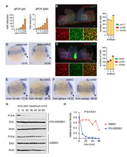
Fgf signaling inhibits endoderm specification (A) qPCR for fgf3 and fgf8a in zebrafish dissociated embryos treated with increasing concentrations of NODAL, mean ± SEM, n >3. (B) Left panel: Flat-mounted 80% epiboly embryo stained for sox32 and sox17. Box with dashed white line indicates the enlarged area presented in the bottom panels where the staining is shown for the individual genes and the two combined. Right panel: quantification of endoderm progenitors from three embryos stained as in left panel. (C) As in (B) but for sox32 and foxa2 in 70% epiboly embryos. The strong foxa2 staining in the center of the flat-mount image corresponds to the foxa2 expressing axial mesoderm. (D) Representative images of either uninjected or fgf8a mRNA-injected embryos (10 pg) isolated at 75% epiboly and stained for sox17 by WISH. Overexpression of fgf8a results in reduced endodermal cell numbers. Arrowhead, dorsal forerunner cells, which mark the most posterior end of the embryo to compare staging. (E) Representative images of 75% epiboly embryos stained for sox17 by WISH after treatment with either DMSO as control or 10 μM SU-5402. Chemicals were added from the 16-cell stage. Arrowhead, dorsal forerunner cells. Note that as well as increasing sox17-positive endoderm progenitors, SU-5402 also partially inhibits cell migration. (F) Same as in (E) but for embryos treated from sphere stage. Note that Fgf signaling inhibition in (E) and (F) both result in more endodermal progenitors. (G) 50% epiboly embryos were treated with DMSO or PD-0325901 for the times shown. Whole cell extracts were Western blotted for P-Erk, Erk1 and Actin as a loading control. (H) The Western blots in (G) were scanned and quantitated using ImageJ. All scale bars are 100 μm. |
Reprinted from Developmental Cell, 44(2), van Boxtel, A.L., Economou, A.D., Heliot, C., Hill, C.S., Long-Range Signaling Activation and Local Inhibition Separate the Mesoderm and Endoderm Lineages, 179-191.e5, Copyright (2017) with permission from Elsevier. Full text @ Dev. Cell