
Fig. S5
- ID
- ZDB-FIG-180608-101
- Publication
- van Boxtel et al., 2017 - Long-Range Signaling Activation and Local Inhibition Separate the Mesoderm and Endoderm Lineages
- Other Figures
- All Figure Page
- Back to All Figure Page
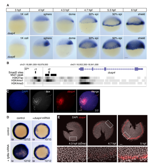
Zebrafish dusp4 is expressed in the first two cell tiers and dephosphorylates Erk (A) WISH for dusp4 and dusp6 expression in 1000-cell (1K) to shield stage embryos. Both dusp4 and dusp6 are initially transiently expressed in a dorsal domain, but from 30% epiboly start to be expressed in the ventral and lateral margin. dusp4 expression remains confined up to the first cell tiers whereas dusp6 is expressed in a much larger domain. The hours post fertilization (hpf) are indicated above. (B) The dusp4 locus from the UCSC genome browser (Zv9) showing the H3K27ac, H3K4me1 and H3K4me3 tracks. The positions of the putative enhancers r1 and r2 are shown, as is the position of the Mixl1 peaks taken from data reported in Nelson et al., 2017. (C) FISH for dusp4 mRNA surrounding Nodal-expressing clone marked with dextran fluorescein (dex). This is the same embryo shown in Figure 5G. (D) WISH for ta after fgf8a and/or dusp4 mRNA injection. Embryos are from the same experiment as the Western blot in Figure 5H. (E) FISH for dusp4 in flat-mounted embryos fixed at the times indicated to show how expression is induced over time from dome stage (4.3 hpf). Note that staining is seen in 2–3 cell tiers from the YSL at the 5 hpf. These are representative embryos from those used to calculate the traces in Figure 5I. All scale bars are 100 μm. |
Reprinted from Developmental Cell, 44(2), van Boxtel, A.L., Economou, A.D., Heliot, C., Hill, C.S., Long-Range Signaling Activation and Local Inhibition Separate the Mesoderm and Endoderm Lineages, 179-191.e5, Copyright (2017) with permission from Elsevier. Full text @ Dev. Cell