
Fig. S7
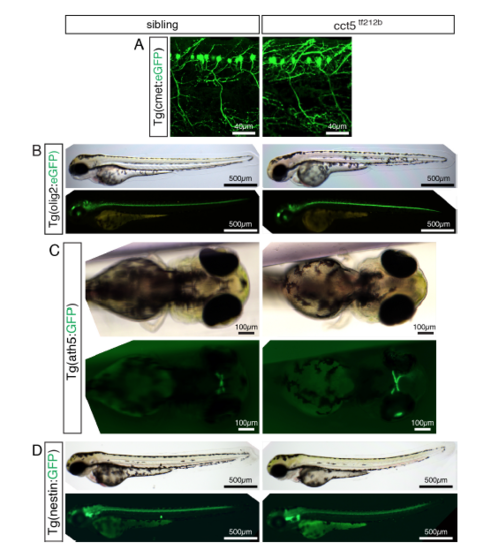
Transgenic marker lines do not reveal a neuronal phenotype within cct5tf212b. Related to Figure 2. (A-D) Representatives of a minimum of 3 analyzed larvae per genotype are shown (n > 3 per genotype). At 3 dpf, no marked phenotype was observed with the following transgenic lines labeling distinct neurons: (A) No marked phenotype of cct5tf212b was observed in the transgenic background of Tg(cmet:eGFP), a marker line for motor neurons (Hall et al., 2007). (B) Similarly, crossing cct5tf212b into the background of Tg(olig2:eGFP) did not reveal a marked phenotype in the oligodendrocytes of cct5tf212b (Shin et al., 2003), documented under bright field and fluorescence conditions. (C) Also crossing cct5tf212b to Tg(ath5:GFP), a marker line for retinal ganglion cells (Masai et al., 2003), did not reveal a neuronal phenotype. (D) Utilizing the Tg(nestin:GFP), which amongst others highlights the neuronal stem cell niche (Kaslin et al., 2009), did not result in a marked phenotype of cct5tf212b mutants. |