
Fig. S5
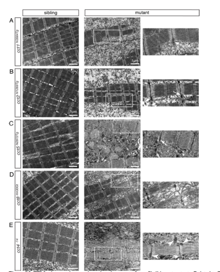
TEM micrographs reveal a reduced amount of myofibril in cct mutants. Related to Figures 1 and 2. (A-E) Sarcomere organization was preserved within all analyzed cct mutants as revealed by EM micrographs of 3-dpf-old larvae. Boxed areas within micrographs of cct mutants are magnified. Arrows point to Z-disk-associated t-tubules and arrowheads notify aggregates. Representatives of a minimum of 3 analyzed larvae per genotype are shown (n > 3 per genotype). (A) The structure of sarcomeres was preserved in cct1hi3564Tg, however the amount of myofibril was reduced in cct1hi3564Tg homozygotes. (B) The reduction in the amount of myofibril and the preserved sarcomere organization was also apparent on TEM micrographs of cct2hi1269Tg. (C) Also in cct5hi2972Tg mutants, sarcomere organization was preserved, but the amount of myofibril was reduced. (D) cct8mn30Gt mutants showed a similar myofibril reduction and unaffected sarcomere organization. (E) In cct4-14 homozygotes the amount of myofibril was reduced and electron-dense aggregates close to Zdisks were documented (arrowheads). |