
Fig. S4
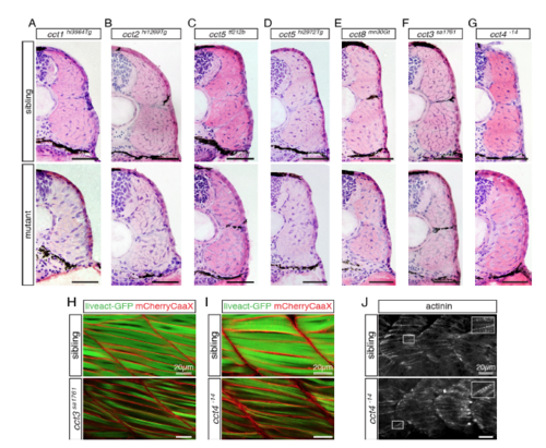
In skeletal muscle, mutations within cct genes can lead to enlarged regions of endomysial connective tissue with reduced amount of myofibril. Related to Figures 1 and 2. (A-G) Cross sections of 3-dpf-old larvae stained for H&E indicate enlarged regions of endomysial connective tissue in cct mutants compared to their siblings. Representatives of a minimum of 6 analyzed larvae per genotype are shown (n > 6 per genotype). (A) Homozygotes of gene trap mutant cct1hi3564Tg and siblings. (B) Homozygotes of gene trap mutant cct2hi1269Tg and siblings. (C) Homozygotes of the mutant cct5tf212b and siblings. (D) Homozygotes of gene trap mutant cct5hi2972Tg and siblings. (E) Homozygotes of gene trap mutant cct8mn30Gt and siblings. (F) Homozygotes of null mutant cct3sa1761 and siblings. (G) Homozygotes of null mutant cct4-14 and siblings. (H and I) Representatives of 4 analyzed larvae per genotype are shown (n = 4 per genotype). (H) The reduced amount of myofibril in cct3sa1761 mutants was confirmed in the transgenic background of Tg(acta1:mCherryCaaX) and Tg(acta1:liveact-GFP). (I) According to the reduced level of birefringence, a reduced amount of myofibril in cct4-14 was visualized by Tg(acta1:mCherryCaaX) and Tg(acta1:liveact-GFP). (J) Regularly spaced actinin-positive Z-bodies of pre-myofibril were detected in siblings as well as in cct4-14 homozygotes. |