
Fig. 1
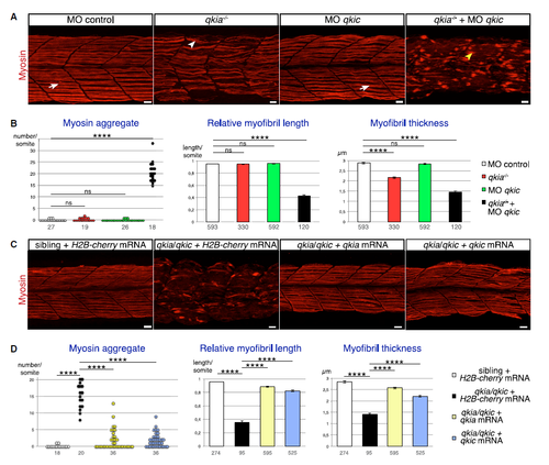
qkia and qkic Functionally Interact to Control Muscle Formation in Slow Muscle Cells (A) qkia and qkic are required for slow muscle formation. Confocal imaging with z projection of myosin-immunostained 26-hpf zebrafish embryos from intercrosses of qkia+/− parents (lateral view). WT or qkia+/− embryos injected with either control MO (left panel, n = 42) or qkic MO (middle right panel, n = 125) exhibit WT myofibrils (arrow). qkia−/− embryos injected with control MO (middle left panel, n = 21) develop moderate myofibril defects (white arrowhead) whereas injection of qkic MO into qkia−/− embryos (right panel, n = 55) leads to a severe disruption of myofibrils (yellow arrowhead). (B) Quantification of the myofibril phenotype illustrated in (A). qkia−/− (n = 5), but not qkic morphant embryos (n = 8) display significantly thinner myofibrils compared with control morphant embryos (n = 9) (Student's t test, p = 2 × 10⁻³⁰ and 0.6, respectively). qkia−/− embryos injected with qkic MO (n = 6) have significantly more myosin aggregate per somite (Student's t test, p = 1.9 × 10⁻¹³), as well as shorter and thinner myofibrils compared with embryos injected with control MO (Student's t test, p = 9 × 10−53 and 1.8 × 10−74, respectively). Numbers under the graph indicate the total number of quantified somites (left graph) and myofibrils (right and middle graphs). ns, not significant. (C) Either qkia or qkic can rescue qkia/qkic myofibril defects. Confocal imaging with z projection of myosin-immunostained 26-hpf zebrafish embryos from intercrosses of qkia+/−;qkic−/− parents (lateral view). qkia−/−;qkic−/− embryos injected with either qkia (n = 10) or qkic (n = 11) mRNA do not develop the severe myofibril defects observed in qkia−/−;qkic−/− embryos injected with H2B-cherry mRNA (n = 12), but rather form myofibrils similar to control (sibling embryos injected with H2B-cherry mRNA, n = 36). (D) Quantification of the myofibril phenotype illustrated in (C). qkia−/−;qkic−/− embryos injected with either qkia (n = 10) or qkic (n = 11) mRNA have significantly fewer myosin aggregate per somite, as well as longer and thicker myofibrils compared with qkia−/−;qkic−/− embryos injected with H2B-cherry mRNA (n = 4) (Student's t test, p < 1 × 10⁻¹⁰ and p < 1 × 10⁻³⁰, respectively). Numbers under the graph indicate the total number of quantified somites (left graph) and myofibrils (right and middle graphs). In all panels, anterior is to the left. Pooled data are presented as mean ± SEM. ∗∗∗∗p < 1 × 10⁻⁴. Scale bars, 10 μm. See also Figure S1. |
| Antibody: | |
|---|---|
| Fish: | |
| Knockdown Reagent: | |
| Anatomical Term: | |
| Stage: | Prim-5 |
| Fish: | |
|---|---|
| Knockdown Reagent: | |
| Observed In: | |
| Stage: | Prim-5 |
Reprinted from Developmental Cell, 42(5), Bonnet, A., Lambert, G., Ernest, S., Dutrieux, F.X., Coulpier, F., Lemoine, S., Lobbardi, R., Rosa, F.M., Quaking RNA-Binding Proteins Control Early Myofibril Formation by Modulating Tropomyosin, 527-541.e4, Copyright (2017) with permission from Elsevier. Full text @ Dev. Cell