
Fig. 5
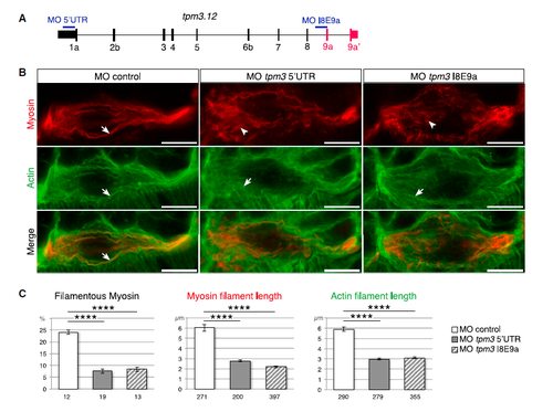
Tropomyosin-3 Is Required for the Early Steps of Myofibril Formation (A) Schematic of the tpm3.12 transcript indicating the position of the two different MO. Exon numbers are indicated below the transcript. (B) Confocal imaging of myosin (red) and actin (green) immunostained embryos at 16 hpf (15-somite stage) (dorsal view from single optical section). WT embryos were injected with control MO or tpm3 MO targeting either the 5′ UTR or the junction between intron 8 and exon 9a (I8E9a junction). In control embryos (n = 31), in somite 7, myosin and actin organize into long filamentous structures (arrows). In both types of tpm3 morphant embryos (5′ UTR MO, n = 14; I8E9a MO, n = 40), actin forms thin filamentous structures (arrows) whereas myosin localizes in short stretches (arrowheads) or remains diffuse. In all panels, anterior is to the left. Scale bars, 10 μm. (C) Quantification of the myofibril phenotype illustrated in (B). In both types of tpm3 morphant embryos (5′ UTR MO, n = 8; I8E9a MO, n = 4), myosin and actin filaments are significantly shorter (Student's t test, 5′ UTR MO: p = 3 × 10⁻²⁰ and 3 × 10⁻²⁷, respectively; I8E9a MO: p = 3 × 10⁻²⁵ and 6 × 10⁻²⁷, respectively) and myosin is less condensed into filaments (Student's t test, 5′ UTR MO: p = 1 × 10⁻⁶; I8E9a MO: p = 1 × 10⁻¹¹) compared with control embryos (n = 4). Numbers under the graph indicate the total number of quantified sections (left graph) and myosin and actin filaments (middle and right graph). Data are presented as mean ± SEM. ∗∗∗∗p < 1 × 10⁻⁴. See also Figure S5. |
| Antibody: | |
|---|---|
| Fish: | |
| Knockdown Reagents: | |
| Anatomical Terms: | |
| Stage: | 14-19 somites |
| Fish: | |
|---|---|
| Knockdown Reagents: | |
| Observed In: | |
| Stage: | 14-19 somites |
Reprinted from Developmental Cell, 42(5), Bonnet, A., Lambert, G., Ernest, S., Dutrieux, F.X., Coulpier, F., Lemoine, S., Lobbardi, R., Rosa, F.M., Quaking RNA-Binding Proteins Control Early Myofibril Formation by Modulating Tropomyosin, 527-541.e4, Copyright (2017) with permission from Elsevier. Full text @ Dev. Cell