
Fig. 3
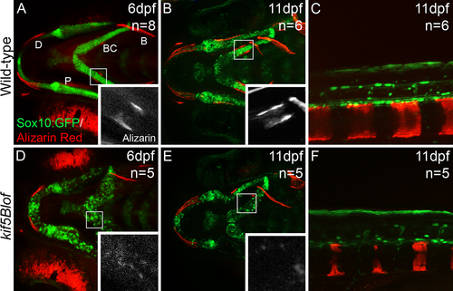
mpeded mineralization in kif5Blof. Confocal projections of images of live embryos at 6 dpf (A, D) and 11 dpf (B, C, E, F). GFP stained chondrocytes and Alizarin Red stained mineralized matrix. Intramembranous bones (MD, P and B) show mineralization but were shorter at 6 dpf (A, D). BC, the only endochondral bone at this stage, mineralization was deficient but evidence of mineralization is detectable (insets in D). At 11 dpf membranous and perichondral ossification had progressed in Wt (B) and vertebraes were apparent (C). In kif5Blof membranous ossification had also progressed (E) and vertebraes were mineralized (F). However, perichondral ossification was defective as indicated by the absence of BC (E). B: branchiostegal; BC: bone collars; P: entopterygoid; D: dentary. |
| Gene: | |
|---|---|
| Fish: | |
| Anatomical Term: | |
| Stage Range: | Day 6 to Days 7-13 |
| Fish: | |
|---|---|
| Observed In: | |
| Stage Range: | Day 6 to Days 7-13 |