
Fig. S3
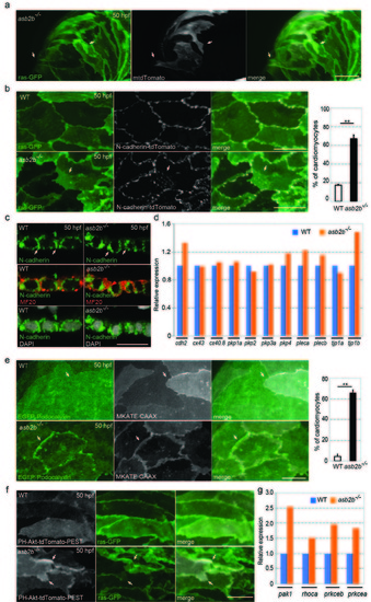
asb2b mutant cardiomyocytes exhibit irregular cell-cell junctions. (a) 3D images of 50 hpf Tg(myl7:ras-GFP) asb2b mutant atria mosaically expressing membrane-tdTomato (mtdTomato). Mosaic expression of mtdTomato was achieved by injecting a myl7:mtdTomato plasmid into Tg(myl7:ras-GFP) asb2b mutant one-cell stage embryos. Membrane protrusions of mtdTomato positive cardiomyocytes are shown (arrows). (b) 3D images of 50 hpf Tg(myl7:ras-GFP);Tg(myl7:N-cadherintdTomato) WT and asb2b mutant atria. N-cadherin-tdTomato molecules in WT cardiomyocytes exhibit lateral localization in cell-cell junctions, while those in asb2b mutants also exhibit localization in overlapping regions (arrows). Number of cardiomyocytes exhibiting N-cadherin-tdTomato punctae in overlapping regions with neighbouring cardiomyocytes in 50 hpf WT and asb2b mutant hearts (n =5 hearts, with averages taken from 25 cardiomyocytes per heart). (c) Co-staining for N-cadherin (green), Myosin heavy chain (MF20, red) and DAPI (white) in 50 hpf WT and asb2b mutant hearts, transverse views. No obvious differences in N-cadherin expression are observed (arrows). (d) Relative mRNA expression (microarray) of junction component genes in isolated 50 hpf WT and asb2b mutant hearts. Gene names can be found in Supplementary Table 1. (e) 3D images of 50 hpf Tg(myl7:EGFP-Podocalyxin);Tg(myl7:MKATE-CAAX) WT and asb2b mutant atria. In WT hearts, EGFP-Podocalyxin shows uniform distribution across cell-cell borders (arrows), while those in asb2b mutants exhibit peripheral localization (arrows). Number of cardiomyocytes exhibiting EGFP-Podocalyxin localization in cardiomyocyte cell-cell borders in 50 hpf WT and asb2b mutant hearts (n=5 hearts, with averages taken from 20 cardiomyocytes per heart). (f) 3D images of 50 hpf Tg(myl7:ras- GFP) WT and asb2b mutant atria expressing PH-Akt-tdTomato-PEST. asb2b mutant cardiomyocytes exhibit enriched localization of PH-Akt-tdTomato-PEST in membrane protrusions (arrows). (g) Relative mRNA expression (microarray) of pak1, rhoca, prkceb and prkcea in isolated 50 hpf WT and asb2b mutant hearts. Gene names can be found in Supplementary Table 1. **P<0.01 by one-way analysis of variance (ANOVA) followed by Tukey’s HSD test. Error bars, SEM. Scale bars, 20 μm. |