
Fig. S1
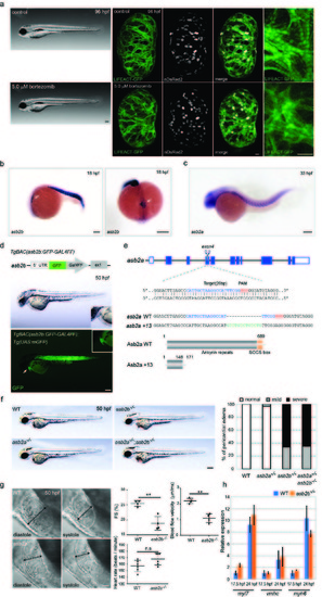
asb2b mutant zebrafish exhibit cardiac defects. (a) Brightfield micrographs of 96 hpf larvae treated with DMSO (control) and bortezomib. Lateral views, anterior to the left. Scale bar, 200 μm. 3D images of 96 hpf Tg(myl7:LIFEACTGFP) hearts in animals treated with DMSO (control) and bortezomib. Animals were treated from 24 until 96 hpf. Scale bar, 20 μm. (b) in situ hybridization for asb2b expression in 18 hpf embryos. Lateral view, anterior to the left; dorsal view, anterior to the bottom. asb2b is expressed in the heart cone (arrows) as well as in the somites. (c) in situ hybridization for asb2a expression in 33 hpf embryos. Lateral view, anterior to the left. Scale bars, 200 μm. (d) Schematic representation of the reporter transgene: a GFPGAL4FF- polyA-Kmr cassette was inserted at the ATG start codon in an asb2b BAC. Lateral views of a 50 hpf Tg(asb2b:GFP-GAL4FF);Tg(UAS:mGFP) embryo and close-up views of the heart; anterior to the left. An arrow points to the heart. GFP is expressed in the heart and somites. Scale bar, 200 μm. (e) Schematic representation of asb2a locus and the gRNA. Target sequence of gRNA and PAM are highlighted in blue and red, respectively. Inserted nucleotides are indicated in green. Predicted structure of WT and Asb2a +13 mutant proteins. asb2a +13 allele is predicted to encode a truncated polypeptide containing 25 incorrect amino acids (147-171 aa). (f) Bright-field micrographs of 50 hpf WT, asb2a mutant, asb2b mutant, and asb2a;asb2b double mutant embryos in lateral views. Scale bar, 200 μm. percentage of WT and mutant embryos exhibiting pericardial edema (n=50 fish). (g) Lateral close-up views of 50 hpf WT and asb2b mutant hearts in diastole and systole. Anterior to the left. Two-headed arrows indicate width of the ventricle. Quantification of ventricular fractional shortening (FS) and aortic blood flow velocity in WT and asb2b mutants. Heart rate measurement in 50 hpf WT and asb2b mutants (n=4 to 6 fish). Scale bar, 20 μm. (h) Relative mRNA expression (qPCR) of myl7, vmhc and myh6 expression at 17.5 and 24 hpf in WT and asb2b mutants (n=2 technical replicates, RNA samples were obtained from 50 embryos). **P<0.01 by one-way analysis of variance (ANOVA) followed by Tukey’s HSD test. Error bars, SEM. |