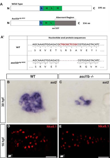
nkx6.1 expression is not repressed by Ascl1b. A Schematic representation of wild-type (WT) and mutant Ascl1b ulg-M2C proteins. The basic domain (b) (+70/+78) is represented by a blue box and the helix loop helix (HLH) domain (+79/+123) by a green box. The coding region of the mutant Ascl1b ulg-M2C protein contains an 11-bp deletion after the aa 107 codon, leading to a frameshift and the production of an aberrant region of 48 aa instead of the second helix, known to be essential for the function of the bHLH proteins. A′ Table showing part of the nucleotide and protein sequence of the ascl1b gene and of the mutated form in the Ascl1b ulg-M2C mutant. B, C WISH showing the drastic reduction of somatostatin expression in the ascl1b -/- mutant compared to the WT embryo at 30 hpf. This phenotype is identical to the one of embryos injected with a translation-blocking morpholino, targeting the translation start site of ascl1b mRNA [15], [77], [78], suggesting that the mutant Ascl1b ulg-M2C is effectively a null mutant. D, E Confocal projection images of Nkx6.1 immunodetection showing equivalent number of nkx6.1+ cells in WT and ascl1b-/- mutants at 16 hpf. All views are ventral with the anterior part to the left. Scale bars = 40 µm.
|
