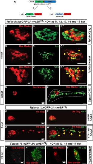
ascl1b-expressing cells give rise exclusively to endocrine cells of both dorsal and ventral bud. a–e′, h–i′ Genetic lineage tracing using the Cre-loxP system.a Schematic representation of the genetic lineage tracing experiments. The transgenic line Tg(ascl1b:eGFP-creER T2 ) was crossed with the Tg(ubi:loxP-eGFP-loxP-mCherry) line, abbreviated Tg(ubi:Switch), or with the Tg(ubi:loxP-AmCyan-loxP-ZsYellow) line, abbreviated Tg(ubi:CSY), treated with 4-hydroxytamoxifen (4OHT) at 11, 12, 13, 14, and 15 hpf (b–e′) or at 13, 14, and 17 dpf (h–i′) and fixed for analysis at the indicated times. Black triangles in a represent loxP sites. b–e′ Immunodetection of CRE-mediated rec markers (ZsYellow or mCherry, red) and Isl1 (b–b"), Ins (c–c"), Gcg (d–d"), or Nkx6.1 (e, e′) in 4OHT-treated embryos (green). The dotted white line delimits the pancreas (e′). Yellow arrows point to cells co-expressing rec marker (ZsYellow or mCherry) and the respective hormones (Ins or Gcg). f–g′ Short-term lineage tracing: immunodetection of GFP and the Ins and Gcg hormones in 5-dpf Tg(ascl1b:eGFP-creER T2 ) embryos treated from 3 to 5 dpf with DMSO (f, f′) or with the Notch signaling inhibitor, LY411575 (g, g′). Yellow arrows in g′ point to GFP+/Ins+/Gcg+ secondary endocrine cells found in the IPDs. h–i′ Immunodetection at 20 dpf of the CRE-mediated rec marker mCherry together with Ins and Gcg in 4OHT-treated larvae. All views are ventral with the anterior part to the left and represent z-plane confocal images (b–d") or confocal projection images (e–i′). Scale bars = 20 µm
|
