
Fig. S3
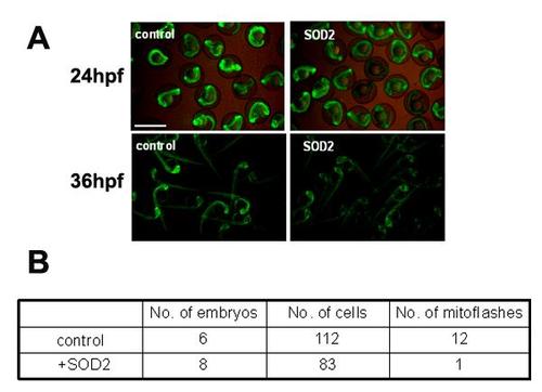
Basal cpYFP signal was decreased in zebrafish embryos overexpressing sod2. Injection of sod2 mRNA decreased the cpYFP fluorescent level at 24 hpf and 36 hpf (Figure A), and suppressed mitoflash activity in red fibers of embryos at 2 dpf (Figure B). Tg(β-actin:mt-cpYFP) transgenic embryos were injected with sod2 mRNA and were then used for measuring mitoflashes in vivo. The red and white fibers of the trunk skeletal muscles were recorded for mitoflashes, and imaged using a 40X, 1.3NA H2O immersion objective at a sampling rate of 1.57 s/frame on Zeiss 710 confocal microscope. Time-lapse images of 100 frames were acquired continuously each time. Scale bar, 10 µm. |