
FIGURE
Fig. S6
- ID
- ZDB-FIG-150831-6
- Publication
- Iyengar et al., 2015 - Poised Regeneration of Zebrafish Melanocytes Involves Direct Differentiation and Concurrent Replenishment of Tissue-Resident Progenitor Cells
- Other Figures
- All Figure Page
- Back to All Figure Page
Fig. S6
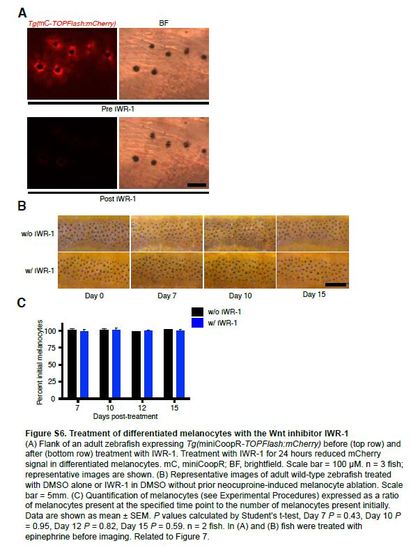
Treatment of differentiated melanocytes with the Wnt inhibitor IWR-1 (A) Flank of an adult zebrafish expressing Tg(miniCoopR-TOPFlash:mCherry) before (top row) and after (bottom row) treatment with IWR-1. Treatment with IWR-1 for 24 hours reduced mCherry signal in differentiated melanocytes. mC, miniCoopR; BF, brightfield. Scale bar = 100 µM. n = 3 fish; representative images are shown. (B) Representative images of adult wild-type zebrafish treated with DMSO alone or IWR-1 in DMSO without prior neocuproine-induced melanocyte ablation. Scale bar = 5mm. (C) Quantification of melanocytes (see Experimental Procedures) expressed as a ratio of melanocytes present at the specified time point to the number of melanocytes present initially. Data are shown as mean ± SEM. P values calculated by Student’s t-test, Day 7 P = 0.43, Day 10 P = 0.95, Day 12 P = 0.82, Day 15 P = 0.59. n = 2 fish. In (A) and (B) fish were treated with epinephrine before imaging. Related to Figure 7. |
Expression Data
Expression Detail
Antibody Labeling
Phenotype Data
Phenotype Detail
Acknowledgments
This image is the copyrighted work of the attributed author or publisher, and
ZFIN has permission only to display this image to its users.
Additional permissions should be obtained from the applicable author or publisher of the image.
Reprinted from Developmental Cell, 33(6), Iyengar, S., Kasheta, M., Ceol, C.J., Poised Regeneration of Zebrafish Melanocytes Involves Direct Differentiation and Concurrent Replenishment of Tissue-Resident Progenitor Cells, 631-43, Copyright (2015) with permission from Elsevier. Full text @ Dev. Cell