
FIGURE
Fig. S5
- ID
- ZDB-FIG-150831-5
- Publication
- Iyengar et al., 2015 - Poised Regeneration of Zebrafish Melanocytes Involves Direct Differentiation and Concurrent Replenishment of Tissue-Resident Progenitor Cells
- Other Figures
- All Figure Page
- Back to All Figure Page
Fig. S5
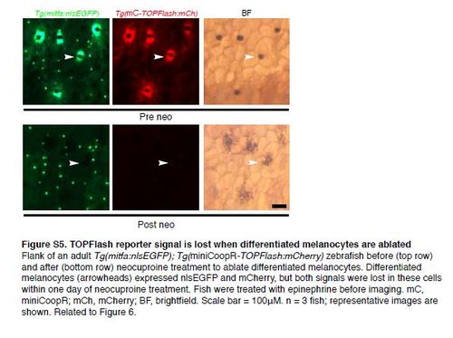
TOPFlash reporter signal is lost when differentiated melanocytes are ablated Flank of an adult Tg(mitfa:nlsEGFP); Tg(miniCoopR-TOPFlash:mCherry) zebrafish before (top row) and after (bottom row) neocuproine treatment to ablate differentiated melanocytes. Differentiated melanocytes (arrowheads) expressed nlsEGFP and mCherry, but both signals were lost in these cells within one day of neocuproine treatment. Fish were treated with epinephrine before imaging. mC, miniCoopR; mCh, mCherry; BF, brightfield. Scale bar = 100µM. n = 3 fish; representative images are shown. Related to Figure 6. |
Expression Data
Expression Detail
Antibody Labeling
Phenotype Data
Phenotype Detail
Acknowledgments
This image is the copyrighted work of the attributed author or publisher, and
ZFIN has permission only to display this image to its users.
Additional permissions should be obtained from the applicable author or publisher of the image.
Reprinted from Developmental Cell, 33(6), Iyengar, S., Kasheta, M., Ceol, C.J., Poised Regeneration of Zebrafish Melanocytes Involves Direct Differentiation and Concurrent Replenishment of Tissue-Resident Progenitor Cells, 631-43, Copyright (2015) with permission from Elsevier. Full text @ Dev. Cell