
Fig. 7
- ID
- ZDB-FIG-150831-13
- Publication
- Iyengar et al., 2015 - Poised Regeneration of Zebrafish Melanocytes Involves Direct Differentiation and Concurrent Replenishment of Tissue-Resident Progenitor Cells
- Other Figures
- All Figure Page
- Back to All Figure Page
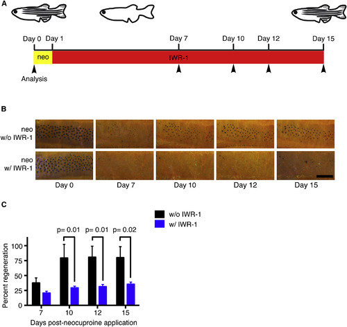
Wnt Signaling Regulates Melanocyte Regeneration (A) Timeline of the experiment. Adult wild-type zebrafish were treated with neocuproine for 24 hr to ablate differentiated melanocytes and were then treated with the Wnt inhibitor IWR-1 for 14 days. Control animals were treated with neocuproine and then DMSO alone. Fish were scored for regeneration melanocytes at 7, 10, 12, and 15 days (arrowheads) after neocuproine treatment. neo, neocuproine. (B) Representative images of control (top row) or IWR-1-treated (bottom row) animals during melanocyte regeneration. Fish were treated with epinephrine prior to imaging. neo, neocuproine. Scale bar, 500 µM. (C) Quantification of melanocyte regeneration. Data are shown as mean ± SEM; p values calculated by Student’s t test, Day 7 p = 0.36. See also Figure S6. |
Reprinted from Developmental Cell, 33(6), Iyengar, S., Kasheta, M., Ceol, C.J., Poised Regeneration of Zebrafish Melanocytes Involves Direct Differentiation and Concurrent Replenishment of Tissue-Resident Progenitor Cells, 631-43, Copyright (2015) with permission from Elsevier. Full text @ Dev. Cell