
FIGURE
Fig. S3
- ID
- ZDB-FIG-150831-3
- Publication
- Iyengar et al., 2015 - Poised Regeneration of Zebrafish Melanocytes Involves Direct Differentiation and Concurrent Replenishment of Tissue-Resident Progenitor Cells
- Other Figures
- All Figure Page
- Back to All Figure Page
Fig. S3
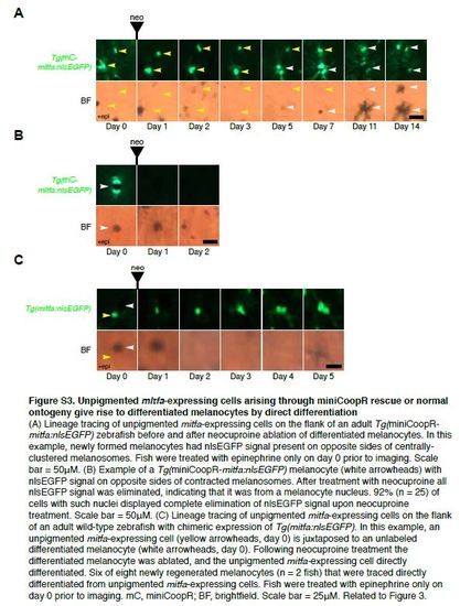
Unpigmented mitfa-expressing cells arising through miniCoopR rescue or normal ontogeny give rise to differentiated melanocytes by direct differentiation (A) Lineage tracing of unpigmented mitfa-expressing cells on the flank of an adult Tg(miniCoopRmitfa: nlsEGFP) zebrafish before and after neocuproine ablation of differentiated melanocytes. In this example, newly formed melanocytes had nlsEGFP signal present on opposite sides of centrallyclustered melanosomes. Fish were treated with epinephrine only on day 0 prior to imaging. Scale bar = 50µM. (B) Example of a Tg(miniCoopR-mitfa:nlsEGFP) melanocyte (white arrowheads) with nlsEGFP signal on opposite sides of contracted melanosomes. After treatment with neocuproine all nlsEGFP signal was eliminated, indicating that it was from a melanocyte nucleus. 92% (n = 25) of cells with such nuclei displayed complete elimination of nlsEGFP signal upon neocuproine treatment. Scale bar = 50µM. (C) Lineage tracing of unpigmented mitfa-expressing cells on the flank of an adult wild-type zebrafish with chimeric expression of Tg(mitfa:nlsEGFP). In this example, an unpigmented mitfa-expressing cell (yellow arrowheads, day 0) is juxtaposed to an unlabeled differentiated melanocyte (white arrowheads, day 0). Following neocuproine treatment the differentiated melanocyte was ablated, and the unpigmented mitfa-expressing cell directly differentiated. Six of eight newly regenerated melanocytes (n = 2 fish) that were traced directly differentiated from unpigmented mitfa-expressing cells. Fish were treated with epinephrine only on day 0 prior to imaging. mC, miniCoopR; BF, brightfield. Scale bar = 25µM. Related to Figure 3. |
Expression Data
Expression Detail
Antibody Labeling
Phenotype Data
Phenotype Detail
Acknowledgments
This image is the copyrighted work of the attributed author or publisher, and
ZFIN has permission only to display this image to its users.
Additional permissions should be obtained from the applicable author or publisher of the image.
Reprinted from Developmental Cell, 33(6), Iyengar, S., Kasheta, M., Ceol, C.J., Poised Regeneration of Zebrafish Melanocytes Involves Direct Differentiation and Concurrent Replenishment of Tissue-Resident Progenitor Cells, 631-43, Copyright (2015) with permission from Elsevier. Full text @ Dev. Cell