
Fig. 10
- ID
- ZDB-FIG-150319-14
- Publication
- Takeuchi et al., 2015 - Establishment of Gal4 transgenic zebrafish lines for analysis of development of cerebellar neural circuitry
- Other Figures
- All Figure Page
- Back to All Figure Page
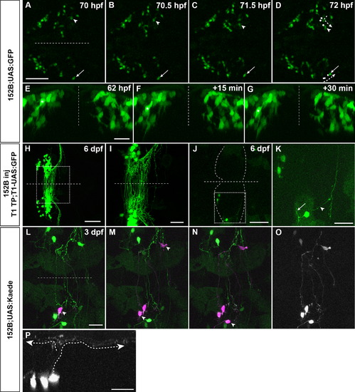
Development and anatomy of granule cells revealed by gSA2AzGFF152B line. (A–G) Dorsal projection views (A–D) and cross section images (E-G) of gSA2AzGFF152B; UAS:GFP larval cerebella. Single cells on the left and right sides (arrowheads and arrows in A–D, asterisks in E–G) are traced during the indicated periods. Trajectories of the marked somata are shown in D (the positions at 70, 70.5, and 71.5 hpf are indicated by white circles and squares). (H–K) Tol1-mediated transgenesis of UAS:GFP reporter in gSA2AzGFF152B line. 5 pg of UAS:GFP reporter DNA in a Tol1 donor vector (T1-UAS:GFP) and 50 pg of Tol1 transposase mRNA were injected into an one-cell stage gSA2AzGFF152B embryo (H, I). 6.6 pg of T1-UAS:GFP DNA and 16.6 pg of Tol1 transposase mRNA (J, K) were injected into one blastomere of a 4-to-8-cell stage gSA2AzGFF152B embryos. GFP expression was detected at 6 dpf. Axon and dendrite structure are indicated by arrowhead and arrow, respectively, in K. (L–O) Combination of mosaic UAS:Kaede and laser irradiation shows the structure of granule cells at a single cell resolution. gSA2AzGFF152B line was crossed with mosaic (partially silenced) UAS:Kaede line. Single somata of granule cells were marked by laser-irradiation one by one at 3 dpf (one in L, three in M, and four in N, marked by arrowheads). Expression of original (green) and photoconverted Kaede (magenta) is shown in L–N. (O, P) Dorsal view and cross section image for photoconverted Kaede in N. Note granule cells in the corpus cerebelli shows typical T-shaped parallel fibers (marked by dotted lines with arrowheads in P). The midlines are indicated by dotted lines. Scale bars: 25 µm in A (applied to A–D); 20 µm in E (applied to E–G); 50 µm in H, J; 20 µm in I, K; 20 µm in L (applied to L–O); 20 µm in P. |
| Genes: | |
|---|---|
| Fish: | |
| Anatomical Term: | |
| Stage Range: | Pec-fin to Protruding-mouth |
Reprinted from Developmental Biology, 397(1), Takeuchi, M., Matsuda, K., Yamaguchi, S., Asakawa, K., Miyasaka, N., Lal, P., Yoshihara, Y., Koga, A., Kawakami, K., Shimizu, T., Hibi, M., Establishment of Gal4 transgenic zebrafish lines for analysis of development of cerebellar neural circuitry, 1-17, Copyright (2015) with permission from Elsevier. Full text @ Dev. Biol.