
Fig. 6
- ID
- ZDB-FIG-150319-10
- Publication
- Takeuchi et al., 2015 - Establishment of Gal4 transgenic zebrafish lines for analysis of development of cerebellar neural circuitry
- Other Figures
- All Figure Page
- Back to All Figure Page
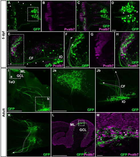
Climbing fibers visualized with the hspGFFDMC28C line. Immunostaining of hspGFFDMC28C; UAS:GFP larvae (A–H) and adult brains (I–M) with anti-GFP (green) and anti-Pvalb7 (magenta) antibodies. CFs are indicated by arrowheads (A, E) or dotted lines (F, Jb, K). (A–C) Dorsal views of the 5-dpf larval hindbrain, with anterior to the left. (D) High magnification view of the box in A. CFs crossing the midline are indicated by arrowheads. (E) Lateral view of the 5-dpf larval hindbrain, with anterior to the left. (F–H) High magnification views of the box in E. (I) Lateral view (with anterior to the left) of the adult brain, subjected to whole mount staining and optical clearing. (Ja, Jb) High magnification views of the boxes in I. (K) Ventral view of the IO region. (L) Sagittal section, with anterior to the left. (M) High magnification view of the box in L. The localization of GFP+ axonal termini on the Pvalb7+ somata of Purkinje cells is indicated by arrowheads. Scale bars: 100 µm in A (applied to A–C); 20 µm in D; 100 µm in E; 20 µm in F (applied to F–H); 400 µm in I; 200 µm in Ja (applied to Ja, Jb, K); 400 µm in L; 20 µm in M. |
| Gene: | |
|---|---|
| Antibody: | |
| Fish: | |
| Condition: | |
| Anatomical Terms: | |
| Stage Range: | Day 5 to Adult |
Reprinted from Developmental Biology, 397(1), Takeuchi, M., Matsuda, K., Yamaguchi, S., Asakawa, K., Miyasaka, N., Lal, P., Yoshihara, Y., Koga, A., Kawakami, K., Shimizu, T., Hibi, M., Establishment of Gal4 transgenic zebrafish lines for analysis of development of cerebellar neural circuitry, 1-17, Copyright (2015) with permission from Elsevier. Full text @ Dev. Biol.