
FIGURE
Fig. 4
- ID
- ZDB-FIG-150115-5
- Publication
- Hartsock et al., 2014 - In vivo analysis of Hyaloid vasculature morphogenesis in zebrafish: A role for the lens in maturation and maintenance of the Hyaloid
- Other Figures
- All Figure Page
- Back to All Figure Page
Fig. 4
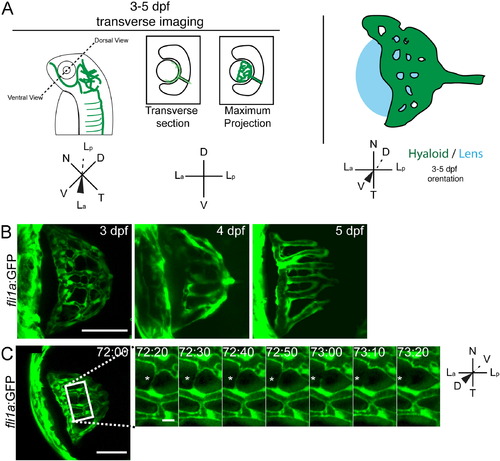
Stage III: refinement of the hyaloid network. (A) Schematic of imaging angles and maximum projections data. Drawings not to scale. (B) Maximum projections of 3-5 dpf hyaloid in transverse imaging view demonstrating the continued reduction and refinement of the vessel network. (C) Still images from time-lapse movies demonstrating the further reduction in vessel complexity as fibers connecting adjacent vessels retract (asterisk). hh:mm. (A,B) Scale bar=50µm; inset scale bar=5 µm. D: Dorsal, V: Ventral, N: Nasal, T: Temporal, La: Lens anterior, Lp: Lens posterior. |
Expression Data
| Gene: | |
|---|---|
| Fish: | |
| Anatomical Term: | |
| Stage Range: | Protruding-mouth to Day 5 |
Expression Detail
Antibody Labeling
Phenotype Data
Phenotype Detail
Acknowledgments
This image is the copyrighted work of the attributed author or publisher, and
ZFIN has permission only to display this image to its users.
Additional permissions should be obtained from the applicable author or publisher of the image.
Reprinted from Developmental Biology, 394(2), Hartsock, A., Lee, C., Arnold, V., Gross, J.M., In vivo analysis of Hyaloid vasculature morphogenesis in zebrafish: A role for the lens in maturation and maintenance of the Hyaloid, 327-39, Copyright (2014) with permission from Elsevier. Full text @ Dev. Biol.