
Fig. 7
- ID
- ZDB-FIG-150115-8
- Publication
- Hartsock et al., 2014 - In vivo analysis of Hyaloid vasculature morphogenesis in zebrafish: A role for the lens in maturation and maintenance of the Hyaloid
- Other Figures
- All Figure Page
- Back to All Figure Page
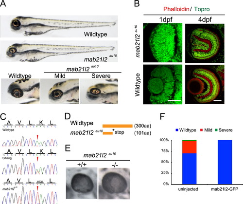
mab21l2au10 mutants possess defects in lens formation. (A) Images of phenotypically wild-type sibling and mab21l2au10 mutants at 4 dpf. High-magnification views of the eyes of mild and severe mab21l2au10 mutants. (B) Transverse cryossections of wild-type and severe mab21l2au10 mutants at 1 and 4 dpf highlighting the lack of a lens in severe mab21l2au10 mutants. Scale bars=50 µm. (C) Genomic sequences from wild-type, heterozygous and mab21l2au10 mutants. mab21l2au10 mutants possess an A->T transversion at position 301, resulting in a premature stop codon at amino acid 101. (D) Schematic of protein length of wild-type and mab21l2au10 mutant. (E) High magnification view of eye in sibling (wild-type) and mab21l2au10 mutant embryo injected with mab21l2-GFP (rescue). (F) Quantification of lens phenotype after mab21l2-GFP injection. |
| Fish: | |
|---|---|
| Observed In: | |
| Stage Range: | Prim-5 to Day 4 |
Reprinted from Developmental Biology, 394(2), Hartsock, A., Lee, C., Arnold, V., Gross, J.M., In vivo analysis of Hyaloid vasculature morphogenesis in zebrafish: A role for the lens in maturation and maintenance of the Hyaloid, 327-39, Copyright (2014) with permission from Elsevier. Full text @ Dev. Biol.