
Fig. 1
- ID
- ZDB-FIG-141007-102
- Publication
- Cardozo et al., 2014 - Cdon acts as a Hedgehog decoy receptor during proximal-distal patterning of the optic vesicle
- Other Figures
- All Figure Page
- Back to All Figure Page
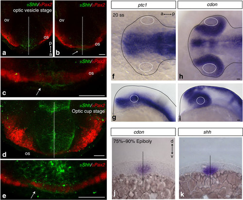
(a–c) Ventral views of anterior zebrafish forebrain at optic vesicle (a is ventral to b) and optic cup (d, e) stages immunostained in toto with antibodies against Pax2 and Shh. (f–i) Dorsal (f,h) and lateral (g,i) views of zebrafish embryos hybridized in toto with probes specific for ptc1 and cdon at the 20 somites’ stage (20 ss). (j–k) Coronal sections of embryos (75–90% epiboly) hybridized for cdon and shh. The white arrows in b,c and e point to Shh immunolabelling in Pax2-positive cells. Dashed lines in a–d,j and k indicate the embryonic midline. Continuous white and black lines in f–i outline the lens and the embryonic border, respectively. a, anterior; d, dorsal; os, optic stalk; ov, optic vesicle; p, posterior; v, ventral. Scale bar, 25 μm. |
| Genes: | |
|---|---|
| Antibody: | |
| Fish: | |
| Anatomical Terms: | |
| Stage Range: | 75%-epiboly to Prim-5 |