
Fig. S4
- ID
- ZDB-FIG-140626-27
- Publication
- Geurtzen et al., 2014 - Mature osteoblasts dedifferentiate in response to traumatic bone injury in the zebrafish fin and skull
- Other Figures
- All Figure Page
- Back to All Figure Page
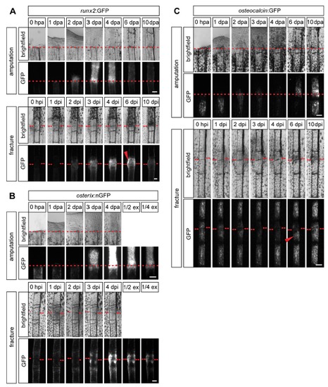
Timing of osteogenesis is similar in fractured versus amputated fin rays, but progression to late osteoblasts is delayed in fractures. (A) Time course of transgene expression in runx2:GFP fins during regeneration after amputation versus repair after fracture. GFP fluorescence is visible from 2 dpa/i in both injury models. runx2:GFP expression is reduced again to levels of uninjured fins at 6 dpa, while it is still high at this time point in fractured fin rays (arrowhead). (B) Time course of transgene expression in osterix:nGFP fins. GFP fluorescence is visible at 3 dpa/i in a similar intensity and is increased at 4 dpa/i in both injury models. ex, exposure. (C) Time course of transgene expression in osteocalcin:GFP fins. GFP fluorescence drops after injury up to 4 dpa/i in both injury models and recurs at 6 dpa in regenerating fins. In fractured fin rays expression levels are equally re-induced at 6 dpi in half of the cases looked at (arrowhead). GFP fluorescence throughout the fractured segment is completely re-established at 10 dpi. dpa, day(s) post amputation. dpi, day(s) post injury. |