
Fig. S1
- ID
- ZDB-FIG-140626-24
- Publication
- Geurtzen et al., 2014 - Mature osteoblasts dedifferentiate in response to traumatic bone injury in the zebrafish fin and skull
- Other Figures
- All Figure Page
- Back to All Figure Page
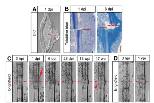
Soft and hard callus formation in fractured fin rays. (A) Longitudinal section view of a fractured fin ray at 1 dpi. The fracture site is indicated by the red dashed line. A swelling covering the fractured hemiray is visible (asterisk). Scale bar, 50 µm. DIC, differential interference contrast. dpi, day post injury. (B) Toluidine blue stained sections of fractured fin hemirays at 1 dpi and 6 dpi. At 6 dpi, collagenous tissue has accumulated at the fracture site (asterisk). Scale bar, 30 µm. (C) Bone fractures in the fin ray heal, however the bone keeps a thickened appearance up to at least 4 months post injury. Live whole mount view of the same fin ray at different time points post fracture. The epidermal thickening forming at 1 dpi is indicated by the arrow. The thickened appearance of the fractured segment is indicated by the arrowhead. Scale bar, 200 µm. hpi, hours post injury. wpi, weeks post injury. (D) The fractured fin ray segment is distinguishable from neighboring unfractured segments at 1 year post injury (ypi). Live whole mount view of the same fin ray at 0 hpi and 1 year later. The thickened bone is indicated by the arrowhead. Scale bar, 200 µm. |