
Fig. 3
- ID
- ZDB-FIG-140527-13
- Publication
- Jiang et al., 2014 - Gene-expression analysis of hair cell regeneration in the zebrafish lateral line
- Other Figures
- All Figure Page
- Back to All Figure Page
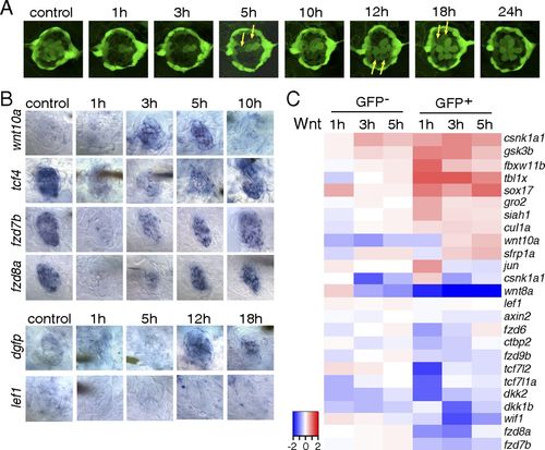
The Wnt/β-catenin pathway is inactive during the early stages of hair cell regeneration. (A) Still images of a Tg(sqET20;sqET4) larval neuromast in the process of neomycin-induced regeneration. All hair cells, except for two immature hair cells, were killed by neomycin by 1 h after neomycin treatment. Two newly formed hair cells (arrows) start to express GFP at 5 h, and other pairs of hair cells (arrows) appear at 12 and 18 h. (B) In situ hybridization of Wnt/β-catenin pathway genes in 5-dpf untreated (control) larvae and in larvae at different time points after neomycin treatment. Expression of wnt10a is increased at 3 and 5 h. Expression of tcf7l2 (tcf4), fzd7b, and fzd8a is largely decreased at 1 h after neomycin. The Wnt/β-catenin reporter line Tg(Tcf/Lef-miniP:dGFP) demonstrates activation starting at 12 h as shown by dgfp expression, but lef1 is not induced in regenerating neuromasts. (C) Heat map of selected Wnt/β-catenin pathway genes based on RNA-Seq results. Log2 ratios of GFP and GFP+ cells at 1, 3, and 5 h after neomycin treatment are color coded (red: up-regulation, blue: down-regulation). Genes that are modulated in GFP cells are neomycin-induced genes. |
| Genes: | |
|---|---|
| Fish: | |
| Condition: | |
| Anatomical Term: | |
| Stage: | Day 5 |