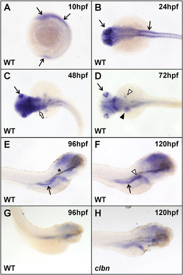
Widespread rnpc3 mRNA expression in early zebrafish embryos becomes more restricted during development. WISH analysis was carried out on clutches of embryos derived from an in-cross of clbnZM heterozygotes using a 599-bp antisense RNA probe designed to hybridize to zebrafish rnpc3 mRNA. (A and B) Widespread rnpc3 mRNA expression in WT embryos at 10 and 24 hpf (arrows). (C and D) From 48 to 72 hpf, rnpc3 mRNA expression becomes progressively restricted to proliferating tissues, including the lens (arrow), pharyngeal region (white arrow), liver and pancreas anlage (black and white arrowheads, respectively), and intestine. (E and F) At 96–120 hpf, rnpc3 mRNA expression is prominent in the digestive organs. Right lateral views show strong expression in the developing pancreas (white arrowhead) and intestine (arrow). (G) Sense probe at 96 hpf produces very weak, nonspecific staining. (H) At 120 hpf, clbn larvae show markedly reduced rnpc3 expression, compared with WT (F). B–D are dorsal views; E–H are right lateral views. Asterisk in E denotes residual pigment after PTU treatment. The same patterns of expression were obtained with a 790-bp probe designed to hybridize to a nonoverlapping region of rnpc3 mRNA. All embryos/larvae shown were genotyped and all images were taken at the same magnification.
|
