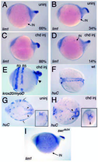
Expansion of lim1+ interneurons depends on Bmp activity. Lateral (A-E,I) and dorsal (F-H) views, anterior is to the left. In 6- to 7-somite uninjected swr/bmp2b homozygotes, 66% show a large expansion in lim1+ interneurons (A) and 34% exhibit a lesser or no expansion in interneurons (B). In the chordin-injected swr/bmp2b mutants, we see a shift to 86% with no or very few (<10) interneurons (C) and 14% that still exhibit normal or expanded numbers (D). Other head and trunk gene expression is unaffected as seen with krox20/myoD expression in a chordin-injected swr/bmp2b embryo (E). The arrowhead points to circular myoD expression in the somitic mesoderm. Expression of huC in 5- to 7-somite wild-type (F), uninjected (G) and chordin-injected (H) swr/bmp2b mutant embryos. (G) Arrows mark the lateral neural cells; (G,H) arrowheads indicate the medial trunk neural cells and insets are posterior views, dorsal to the top, showing a loss of lateral neural cells in ventral regions of injected mutants, while medial neural cells remain. (I) Expansion of lim1+ interneurons in swr/bmp2btdc24 presumptive null mutants.
|
