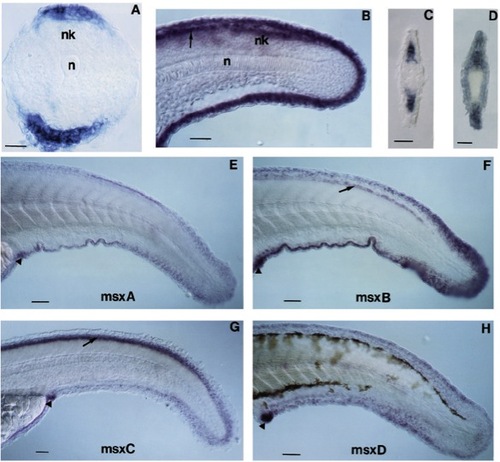
Cells in the median fin fold express msx genes. (A) Transverse section through the tailbud of a 16 h zebrafish embryo. The dorsal side is to the top. Cells on the median edge of the tail bud express msxB. (B) At 24 h, transcripts of the msxB genes are uniformly distributed along the median fin fold. At 36 h, the most distal cells of the median fin fold and cells that occupy an immediately more proximal position express msxA, msxB and msxD (E, F and H, respectively). In contrast, MsxC expression at 36 h is restricted to cells of the median fin fold that occupy a position immediately proximal to the most distal cells (G). (C,D) Transverse sections through the tail of 48 h zebrafish embryos: msxC expression is confined to the mesenchymal cells of the fin fold (C), msxD transcripts are also found in the mesenchymal cells as well as in the ectodermal cells (D). The arrows indicate cells of the dorsal spinal cord and overlying ectoderm that also express msxB (B,F) and msxC (G). Cells in or around the vent also express all four msx genes (arrowheads in E-H). Embryos in (E-G) were treated with 0.2 mM phenylthiocarbamide to inhibit pigmentation. nk, neural keel; n, notochord. Scale bar: 20 μm in (A,C,D), 40 μm in (B,E-H).
|
