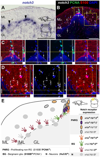
notch3 expression in the adult cerebellar niche.
Cross-sections at the indicated level through the mesencephalon; cerebellar area shown in the micrographs is indicated in the cross-section schematic. A, Brightfield image shows notch3 expressing cells in the cerebellum (black arrowheads). B-D, Confocal images showing localization of the glial marker S100β (red) and PCNA (green), with notch3 by FISH (white). B-C, notch3 is weakly expressed in a small subset of PCNA+ cells in the stem cell niche (unfilled arrowhead). B–D, Strong notch3 expression is detected in S100β+ cells indicated by the white arrows; notch3 expression is also detected in some scattered cells of the ML, IML and GL that do not localize with the analysed markers (unfilled arrows); E, Summary of the expression pattern and cellular characteristics of Notch receptor expressing cells in the adult cerebellum. Abbreviations: GL, granule cell layer; IML, intermediate layer; ML, molecular layer. Scale bars = 50 μm in A and B; 20 μm in C and D.
|
