
Fig. S6
- ID
- ZDB-FIG-140114-7
- Publication
- de Oliveira-Carlos et al., 2013 - Notch receptor expression in neurogenic regions of the adult zebrafish brain
- Other Figures
- All Figure Page
- Back to All Figure Page
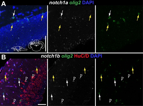
notch1a and notch1b expression in olig2+ and HuC/D + cells of the optic tectum. Confocal images of double FISH showing the localization of notch1a/1b (white), olig2 (green) and HuC/D (red) in the superficial layer of the optic tectum. Cross-sections at the indicated level through the mesencephalon; tectal area shown in the micrographs is indicated in the cross section schematic in A. A–B, notch1a and notch1b are expressed in a subpopulation of olig2 cells (white arrows); yellow arrows indicate Notch receptor - /olig2 + cells. B, notch1b is also expressed in olig2 - /Hu + cells (unfilled white arrowheads). Scale bars = 50 μm. |