
Fig. 6
- ID
- ZDB-FIG-130807-28
- Publication
- Shin et al., 2012 - Intrinsic and extrinsic modifiers of the regulative capacity of the developing liver
- Other Figures
- All Figure Page
- Back to All Figure Page
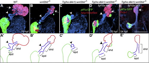
Both the liver and the extrahepatic duct form in Wnt/β-catenin signaling deficient embryo slacking Fgf10a function. Tg(hs:dkk1-GFP);wnt2bb-/- embryos were injected with fgf10a MO, heat-shocked at 18 hpf, and examined at 54 (E) or 70 (D) hpf. The extrahepatic duct was revealed by mAb 2F11 staining, the exocrine pancreas by Tg(ptf1a:GFP) expression, and the liver by Tg(fabp10:RFP) (A-D) or Prox1 (E) expression. Both the liver (arrows) and the extrahepatic duct (ehd) failed to form in Wnt/β-catenin signaling deficient embryos (C), whereas they did form when these embryos were injected with fgf10a MO (D and E). The gallbladder (arrowheads) formed in the extrahepatic duct when the liver formed, whereas it formed in the extrapancreatic duct (epd) when the liver failed to form (C). (A′-E′) The liver, the extrahepatic and extrapancreatic ducts as well as the pancreas are schematically illustrated. Ventral views, anterior up. Scale bar, 100 µm. |
| Genes: | |
|---|---|
| Antibodies: | |
| Fish: | |
| Condition: | |
| Knockdown Reagent: | |
| Anatomical Terms: | |
| Stage Range: | Long-pec to Pec-fin |
| Fish: | |
|---|---|
| Condition: | |
| Knockdown Reagent: | |
| Observed In: | |
| Stage Range: | Long-pec to Pec-fin |
Reprinted from Mechanisms of Development, 128(11-12), Shin, D., Weidinger, G., Moon, R.T., and Stainier, D.Y., Intrinsic and extrinsic modifiers of the regulative capacity of the developing liver, 525-535, Copyright (2012) with permission from Elsevier. Full text @ Mech. Dev.