
Fig. 2
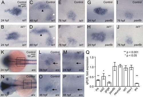
Unchanged expression of early endocrine markers in isl1 mutants. Whole mount in situ hybridizations for isl1 (A–F) and pax6b (G–J) mRNA in control (A, C, E, G, I) and in isl1-/- embryos (B, D, F, H, J) at 24 hpf (A, B, G, H), 48 hpf (C, D) and 76 hpf (E, F, I, J). Note that the pattern and intensity of isl1 signals in the pancreatic mesenchyme (black arrow head) and in the endocrine islet (white arrow head) is indistinguishable in control and mutant embryos. Embryos are shown from ventral with anterior to the left. (K–P) Whole mount in situ hybridizations for arx. (K, N) Dorsal view of control embryos at 24 hpf (K) and 48 hpf (N). Insets mark the position of the pancreatic area shown in higher magnification panels (L M, O, P). Images show a ventral view of control (L, O) and isl1-/- mutant (M, P) embryos. (Q) qPCR analysis for hormone and transcription factor encoding mRNA of isolated pancreata from 96 hpf old embryos. Shown are relative expression levels in pancreata of mutant as compared to control embryos, normalized to the relative expression levels of ins obtained for 96 hpf whole embryonic mRNA. |
| Genes: | |
|---|---|
| Fish: | |
| Anatomical Terms: | |
| Stage Range: | Prim-5 to Protruding-mouth |
Reprinted from Developmental Biology, 378(1), Wilfinger, A., Arkhipova, V., and Meyer, D., Cell type and tissue specific function of islet genes in zebrafish pancreas development, 25-37, Copyright (2013) with permission from Elsevier. Full text @ Dev. Biol.