|
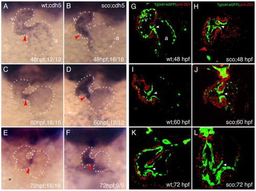
Endocardial cell adhesion and tight junctions were ectopically expressed in the AVC of scote382 mutant hearts. (A–F) Endocardial cell adhesion gene cdh5/ve-cadherin was analyzed by in situ hybridization in wild-type embryos at 48hpf (A), 60hpf (B) and 72hpf (C); and scote382 mutant embryos at 48hpf (D), 60hpf (E) and 72hpf (F). Note gradual restriction of cdh5 in the AVC in wild-type embryos and ectopic expression of cdh5 in the ventricle of scote382 mutant embryos. Red arrowheads point to the AVC. (G–L) Wild-type and scote382 mutant Tg(kdrl:EGFP) transgenic embryos on vibratome sections were subjected to immunostaining with anti-ZO1 antibody. Note that ZO1 was expressed in the AVC of wild-type hearts at 48hpf (G) and downregulated at 60 (I) and 72hpf (K), but remained in the AVC of scote382 mutant hearts at 48 (H), 60 (J) and 72hpf (L). a, atrium; v, ventricle; arrowhead points to the AVC. Dotted lines outline the heart.
|