
Fig. S6
- ID
- ZDB-FIG-130624-9
- Publication
- Wang et al., 2013 - Genetic Interaction between pku300 and fbn2b Controls Endocardial Cell Proliferation and Valve Development in Zebrafish
- Other Figures
- All Figure Page
- Back to All Figure Page
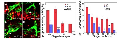
Endocardial cell proliferation is defective in scote382 mutants. (A-E) wild-type (A, C) and scote382 mutant (B, D) Tg(kdrl:eGFP) embryos at 23 hpf and 25 hpf (A, B), 27 hpf and 30 hpf (C, D) were subjected to immunostaining with anti-pH3 antibody. Images were photographed under Zeiss 510 confocal microscope. White arrowheads point to pH3-positive Tg(kdrl:eGFP) endocardial cells in the heart tube. (E) The pH3-positive endocardial cells were scored and statistically analyzed in wild-type and scote382 mutant embryos from 23 to 30 hpf. (F) The BrdU-positive Tg(kdrl:eGFP) endocardial cells were scored and statistically analyzed in wild-type and sco mutant embryos from 23 to 33 hpf. Note that pH3- and BrdU-positive endocardial cells were gradually decreased in scote382 mutants. (E-F) n=3-5; mean±SEM; student’s t-test. |