
Fig. 2
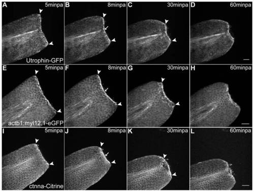
Abrupt tissue contraction and actomyosin cable formation initiate wound healing. A–D Sequential images of a representative in vivo 55 min time-lapse movie of an Utrophin-GFP mRNA-injected 2 dpf larva at different time-points after amputation. A At 5minpa, there is accumulation of actin at the leading edge cells. B By 8 minpa the actin cable is assembled (arrow) and there is tissue contraction - note displacement of arrowheads in comparison with A. C At 30 minpa, the tissue has fully contracted. D At 60 minpa, the wound inflicted by the amputation is closed. E–H Sequential images of a representative in vivo 55 min time-lapse movie of a 2 dpf actb1:myl12.1-eGFP transgenic larva. E At 5minpa, there is accumulation of myosin at the leading edge cells. F By 8 minpa the myosin cable is assembled (arrow) and there is tissue contraction - note displacement of arrowheads in comparison with E. G At 30 minpa, the tissue has fully contracted. H At 60 minpa, the wound inflicted by the amputation is closed. I–L Sequential images of a representative in vivo 55 min time-lapse movie of a 2 dpf ctnna-Citrine transgenic larva at different time-points after amputation. I At 5minpa, there is accumulation of alpha-catenin at the leading edge cells. J By 8 minpa this accumulation appears to localize to the actomyosin cable (arrow) and there is tissue contraction - note displacement of arrowheads in comparison with I. K At 30 minpa, the tissue has fully contracted and alpha-catenin is still localized at the wound edge. L At 60 minpa, the wound inflicted by the amputation is closed. Anterior is on the left and scale bars correspond to 50 μm in all images; n = 5 larvae per condition. |