
Fig. S11
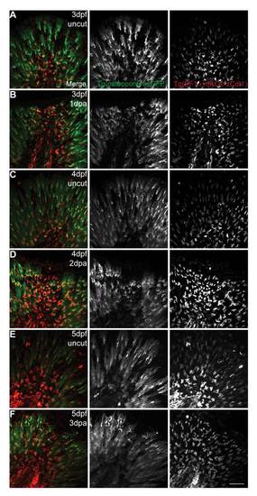
The mesenchymal cells are maintained in G0-G1 phases of the cell cycle regardless of an amputation. Live imaging representative images of double transgenic EF1α:mKO2-zCdt1;osteopontin:eGFP larvae during several stages of the regenerative process and their respective controls. A,C,E are uncut (3 dpf, 4 dpf and 5 dpf respectively) and age matched controls for B,D,F (3 dpf 1 dpa, 4 dpf 2 dpa, 5 dpf 3 dpa respectively). Merged and single color images corresponding to osteopontin:eGFP labeling the cytoplasm of the mesenchymal cells (green) and mKO2-zCdt1 labeling the nuclei of fin fold cells in G0–G1 phases of the cell cycle (red). 3 larvae per condition. Scale bar corresponds to 50 μm. |