
FIGURE
Fig. S6
Fig. S6
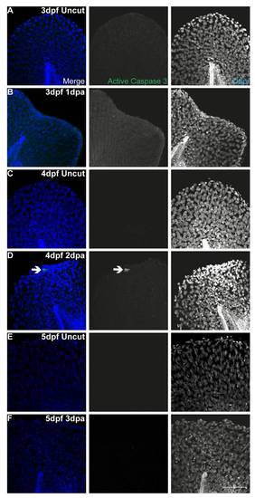
Apoptosis is not present during fin fold regeneration. Representative immunofluorescence with anti-active Caspase3 antibody in uncut and amputated larvae of 3 dpf (A–B), 4 dpf (C–D) and 5 dpf (E–F). Arrow indicates the presence of an apoptotic cell. n = 5 larvae per condition. Scale bar corresponds to 50 μm in all images. |
Expression Data
Expression Detail
Antibody Labeling
Phenotype Data
Phenotype Detail
Acknowledgments
This image is the copyrighted work of the attributed author or publisher, and
ZFIN has permission only to display this image to its users.
Additional permissions should be obtained from the applicable author or publisher of the image.
Full text @ PLoS One