
Fig. S6
- ID
- ZDB-FIG-120720-81
- Publication
- Zou et al., 2012 - Crb Apical Polarity Proteins Maintain Zebrafish Retinal Cone Mosaics via Intercellular Binding of Their Extracellular Domains
- Other Figures
- All Figure Page
- Back to All Figure Page
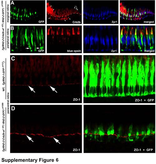
In Tg(RH2-2:Crb2b-sf-EX / RH2-2:GF) pt108b transgenic line, Crb2b-sf-EX and GFP is predominantly expressed in blue cones, and the overall integrity of the OLM is largely unaffected by the transgenic expression, related to Figure 7. A. At the marginal zone, exclusive endogenous Crb2b expression is indicated by the arrows. The lack of GFP and ectopic Crb2b immunoreactivity at the marginal zone suggests that the expression of the transgenes was slightly delayed compared to the endogenous crb2b gene, consistent with the activation pattern of the RH2-2 promoter (Takechi and Kawamura, 2005). The dashed line indicates the location of the OLM, and arrowheads indicate ectopic accumulation of Crb2b-sf-EX. B. The immunohistochemical analyses with anti-blue opsin antibodies (Vihtelic et al., 1999) demonstrated that the transgenes were mainly expressed in the blue cones in the peripheral, paracentral, and central retina in adult Tg(RH2-2:Crb2b-sf-EX / RH2-2:GF) pt108b. Arrowheads indicate GFP expression in some rods, whose cell nuclei were positioned basal to the OLM. C, D. Like in wildtype Tg(RH2-1:GFP) pt112 fish (C), overall integrity of the OLM (visualized by ZO-1 staining, arrows) was largely unaffected in Tg(RH2-2:Crb2b-sf-EX / RH2-2:GFP) pt108b transgenic line (D) at 24 mpf. |
Reprinted from Developmental Cell, 22(6), Zou, J., Wang, X., and Wei, X., Crb Apical Polarity Proteins Maintain Zebrafish Retinal Cone Mosaics via Intercellular Binding of Their Extracellular Domains, 1261-1274, Copyright (2012) with permission from Elsevier. Full text @ Dev. Cell