
Fig. 7
- ID
- ZDB-FIG-120720-48
- Publication
- Zou et al., 2012 - Crb Apical Polarity Proteins Maintain Zebrafish Retinal Cone Mosaics via Intercellular Binding of Their Extracellular Domains
- Other Figures
- All Figure Page
- Back to All Figure Page
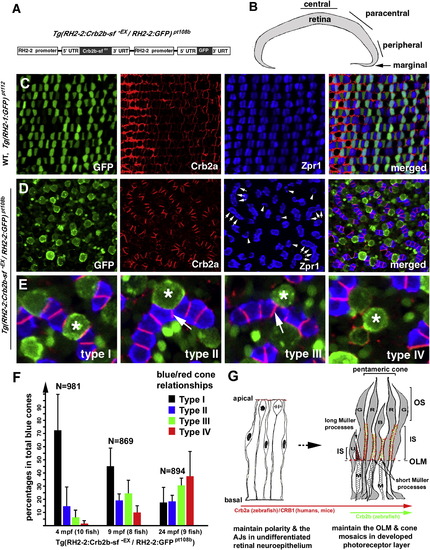
Transgenic Expression of a Secreted Form of the Extracellular Domain of Crb2b-sf (Crb2b-sf-EX) Disrupts Cone Mosaics (A) A schematic illustrates the tandem arrangement of the Crb2b-sf-EX and GFP transgenes driven by the RH2-2 green opsin promoter (Tsujimura et al., 2007). (B) The central, paracentral, peripheral, and marginal regions of the retina are indicated. (C) Photoreceptors are organized in regular mosaic in the paracentral retina of wild-type Tg(RH2-1:GFP) pt112 fish (Zou et al., 2010) at 24 months postfertilization (mpf). (D) Transgenic expression of Crb2b-sf-EX in blue cones disrupted photoreceptor mosaics in the paracentral retina in Tg(RH2-2:Crb2b-sf-EX/RH2-2:GFP)pt108b at 24 mpf. Some double cones were dissociated into solitary Zpr1-positive cells (arrowheads); other double cones abnormally aligned into chains (arrows). (E) In Tg(RH2-2:Crb2b-sf-EX/RH2-2:GFP)pt108b, blue cones (asterisks) were found in four different spatial relationships with red cones: type I, sandwiched between red cones; type II, partially expelled from the red cone sandwich; type III, completely expelled from the red cone sandwich but still attached to the side of the red cones; type IV, solitary without adhering to any Zpr1-positive cones. The arrows indicate the de novo junctions formed between two presumable red cones. (F) Blue cone-secreted Crb2b-sf-EX gradually compromised the association between blue and red cones, resulting in an increase in the percentages of dissociated blue cones over time. Error bars = SD. (G) A diagram summarizes the temporal and spatial expression patterns and functions of Crb proteins during the vertebrate retinal development. Zebrafish Crb2a and its mammalian equivalent CRB1 are broadly expressed and regulate the polarity and integrity of both the undifferentiated neuroepithelium and the developed photoreceptor layer. Crb2b homologs are likely more restrictively expressed in certain types of photoreceptors in a subgroup of vertebrates, such as fish. The restricted expression of Crb2b at the inner segments between green (G), red (R), and blue (B) cones may underlie their structural assembly into pentameric cones in zebrafish; consequently, Crb2b plays a critical role in maintaining cone mosaics. The long apical processes of Müller cells (M) cluster around the UV cones (U), likely preventing them from adhering to pentameric cones. The apical processes of Müller cells between the members of pentameric cones are very short and are thus no impediment to their inner segment associations. See also Figure S6. |
| Gene: | |
|---|---|
| Fish: | |
| Anatomical Terms: | |
| Stage: | Adult |
| Fish: | |
|---|---|
| Observed In: | |
| Stage: | Adult |
Reprinted from Developmental Cell, 22(6), Zou, J., Wang, X., and Wei, X., Crb Apical Polarity Proteins Maintain Zebrafish Retinal Cone Mosaics via Intercellular Binding of Their Extracellular Domains, 1261-1274, Copyright (2012) with permission from Elsevier. Full text @ Dev. Cell