
Fig. S3
- ID
- ZDB-FIG-120720-72
- Publication
- Zou et al., 2012 - Crb Apical Polarity Proteins Maintain Zebrafish Retinal Cone Mosaics via Intercellular Binding of Their Extracellular Domains
- Other Figures
- All Figure Page
- Back to All Figure Page
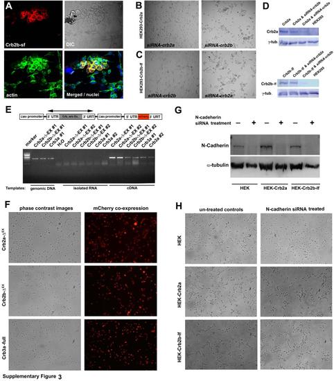
Expression of Crb variants that bear the extracellular domains in HEK cells prompted cell aggregation, related to Figure 4. Expression of full-length Crb2a, Crb2b-sf, or Crb2b-lf in HEK239 cells specifically introduced cell aggregation, and siRNA-mediated suppression of Crb2a or Crb2b-lf abolished cell aggregation. In addition, Crb2a- or Crb2b-lf-mediated HEK293 cell aggregation requires their extracellular domains, but is independent of N-cadherin. A. Transient expression of Crb2b-sf prompted cell aggregation in HEK293 cells. Crb2b-sf positive cells appeared round and formed a tighter and clumping cluster compared to their neighboring non-transfected cells. The cell nuclei were staining with DAPI in blue. B. The aggregation of the Crb2a-expressing HEK293 cells was blocked with the siRNAs against Crb2a but not with the siRNAs against Crb2b. C. The aggregation of the Crb2b-lf-expressing HEK293 cells was blocked with the siRNAs against Crb2b but not with the siRNAs against Crb2a. D. Western blot analyses showed that the protein expression level of Crb2a and Crb2b-lf was specifically and significantly suppressed with the siRNAs. The -tubulin (-tub.) blotting served as loading controls. The cell lines and siRNA treatments were indicated on top of the blots. E. A schematic illustrates the structures of transgenic constructs, each of which contains a gene for Crb2a-ΔEX, Crb2b-ΔEX, or Crb3a as well as a reporter gene of mCherry. Both genes are driven ubiquitously by the CMV promoter. The double-headed arrow indicates the region amplified by RT-PCR reactions, which verified that the Crb genes were expressed in the stably transfected HEK293 cell lines. Two stable clones of each transfection were analyzed by RT-PCR analyses using three different templates: with genomic DNA as positive controls for the robustness of the PCR reactions, with isolated RNA or water as negative controls for genomic DNA contamination, and with cDNA to confirm Crb gene expression. F. Stably transfected monoclonal HEK293 cells that express Crb2a-ΔEX, Crb2b-ΔEX, or Crb3a grew mostly individually. The mCherry expression verified that all cells were transgenic. G. To suppress N-cadherin expression, wildtype HEK293, Crb2a- and Crb2b-lf-expressing HEK 293 cells were transfected with the N-cadherin siRNAs. The expression levels of N-cadherin and cell aggregation phenotypes were analyzed one day after siRNA transfection. Western blotting analyses demonstrated that the N-cadherin siRNA treatments of the three cell lines almost completely abolished their N-cadherin expression. The loading controls were blotted with anti-α-tubulin antibody. H. Compared with untreated HEK293 cells, suppression of N-Cadherin expression in wildtype HEK293 cells reduced their tendency to aggregate. Suppression of N-Cadherin in Crb2a-expressing and Crb2b-lf-expressing HEK293 cells did not abolish the cell aggregation, suggesting the Crb2 can sufficiently mediate cell-cell adhesion independent of N-Cadherin. |
Reprinted from Developmental Cell, 22(6), Zou, J., Wang, X., and Wei, X., Crb Apical Polarity Proteins Maintain Zebrafish Retinal Cone Mosaics via Intercellular Binding of Their Extracellular Domains, 1261-1274, Copyright (2012) with permission from Elsevier. Full text @ Dev. Cell