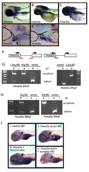
Expression and functional analysis of zebrafish Hox3 genes. (A-C) Expression of PG3 Hox genes in 72 hpf zebrafish embryos. Whole-mount in situ hybridization with 72 hpf embryos showing the expression patterns of Hoxa3a (A), Hoxb3a (B), and Hoxd3a (C). These three PG3 Hox genes were expressed in similar patterns. Arrows indicate expression in the pharyngeal arch region. Sagittal sections of Hoxa3a (D) and Hoxb3a (E) whole-mount in situ hybridization embryos. Hoxa3a and Hoxb3a were both detected to be expressed in the pharyngeal arch mesenchyme and pouch endoderm. Arrows indicate the third pouch. Anterior is to the left, dorsal is up. (F-I) Early knockdown of Hoxa3a and Hoxb3a by the injection of MOs does not affect later thymus development in zebrafish. (F) Scheme of prespliced mRNA of Hoxa3a and Hoxb3a. The red bars indicate the MO specific to the splicing donor or acceptor site. The arrows represent the primers used in RTPCR. (G) RT-PCR products were run on a 1.0% agarose gel; 3.3 ng/embryo or 5 ng/embryo of Hoxa3a MOs (donor and receptor) or mismatched MO were injected into one-cell stage embryos. At 24 hpf, the embryos were collected and analyzed for mRNA splicing. On the gel, the lower bands represent the RT-PCR product of postspliced mRNAand the higher bands represent the product of prespliced mRNA. In the MO-injected embryos, most of the mRNA was prespliced, indicating that the MOs suppressed splicing of target genes effectively at this time point. Hoxa3a (Left) and Hoxb3a (Right) are shown. RT+, PCR with first-strand cDNA; RT, PCR with non-RT control. (H) Hoxa3a RNA splicing in 24 hpf and 48 hpf morphants. At 24 hpf, there was no postspliced mRNA product in the morphants, but at 48 hpf, the postspliced mRNA product was restored. (I) Whole-mount in situ hybridization analysis on Rag-1 expression in the Hoxa3a/Hoxb3a morphants at 1 week. (i) Rag-1 expression was detected in the embryos injected with mismatched MO. (ii) Embryos injected with 5 ng/embryo of Hoxa3 MO specific to splicing donor site showed normal Rag-1 expression. (iii) A total of 5 ng of MOs that were specific to Hoxa3a splicing donor site and acceptor site were coinjected into the embryos. Rag-1 expression in these embryos was not changed. (iv) A total of 3.3 ng/embryos of Hoxb3a MO (splicing donor site) was coinjected with Hoxa3a MOs. Rag-1 expression in these morphants was normal compared with control. Arrowheads indicate Rag-1 expressing cells.
|
