
Fig. 5
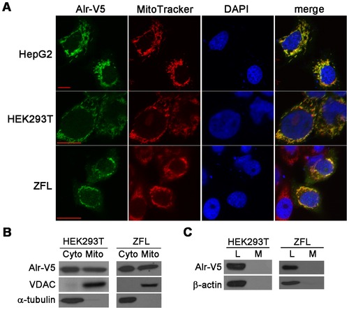
Zebrafish Alr is localized in both the cytosol and mitochondria, but neither in the nucleus nor secreted outside of the cell. A. Alr subcellular localization by immunofluorescent staining. Human hepatocellular carcinoma cells HepG2, human embryonic kidney cells HEK293T and zebrafish liver cells ZFL were transfected with pEF6/V5-His-TOPO plasmid expressing Alr-V5. MitoTracker was used to label the mitochondria and the cells were counter stained with DAPI to mark the nucleus. The Alr protein is co-localized with MitoTracker in the mitochondria, but not present in nucleus. Scale bar is 10 μm. B. Alr subcellular localization by cell fractionation. Western blot revealed that Alr was localized in both the cytosol and mitochondria fractions in transfected HEK293T cells and zebrafish liver cell line (ZFL). Alr was detected by anti-V5 antibody. The mitochondrial porin voltage-dependent anion channel (VDAC) was used as the mitochondria marker while α-tubulin was used as the cytosolic marker. C. Alr was not secreted outside of cell. Alr-V5 was detected in cell lysates but not in the conditioned medium in both HEK293T and ZFL cells. β-actin was used as loading control. L, cell lysate; M, medium. |