
Fig. S6
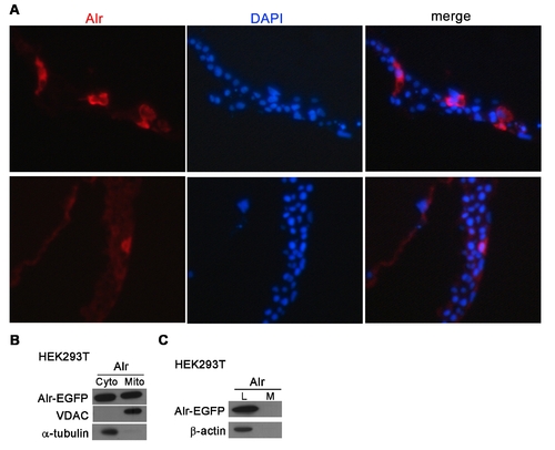
Cellular localization of Alr-EGFP in zebrafish embryo and cultured cells. A. Alr-EGFP is mainly localized in the cytoplasm in zebrafish embryo. The plasmid expressing Alr-EGFP fusion protein under the CMV promoter, was injected into zebrafish 1-cell stage embryos and these embryos were fixed at shield stages (6 hpf) and processed for sectioning. The cryo-sections were stained with mouse anti-GFP primary antibody and Alexa Fluor 568 conjugated anti-mouse IgG secondary antibody. DAPI was used to stain nucleus. Red color shows the predominant presence of Alr-EGFP fusion protein in cytoplasm, but not nucleus. B. Alr-EGFP is localized in both the cytosol and mitochondria. HEK293T cells were transfected with Alr-EGFP expressing plasmid. Cell fractionation followed by Western blot using anti-EGFP antibody revealed that Alr-EGFP was localized in both the cytosol and mitochondria in transfected HEK293T cells. The mitochondrial porin voltage-dependent anion channel (VDAC) was used as the mitochondria marker while α-tubulin was used as the cytosolic marker. C. Alr was not secreted outside of cell. Alr-EGFP is detected by anti-GFP antibody Western blot. The β-actin was used as loading control. L, cell lysate; M, conditioned medium. |