
Fig. 1
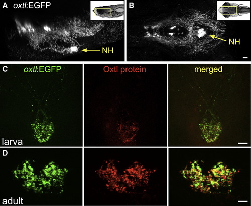
Oxytocinergic Reporter Transgene Tags HN Projections (A and B) 3D reconstruction of the hypothalamic-neurohypophseal system of an optically sectioned 9-day-old zebrafish embryo carrying the oxtl:EGFP transgene (anterior to the left; A, lateral view; B, ventral view). Axonal projections to the neurohypophysis (NH) are highly visible in oxtl:EGFP transgenic animals due to extensive arborization of NH nerve termini. (C and D) Immunohistochemical analysis of Oxytocin protein in oxtl:EGFP transgenic zebrafish showing colocalization of EGFP+ and Oxytocin+ nerve termini in the neurohypophysis of either 6-day-old larva (C, anterior up) or coronally sectioned adult (D); 14-day-old). Scale bars, 20 μm. See also Figure S1 and Movie S1. |
| Genes: | |
|---|---|
| Antibody: | |
| Fish: | |
| Anatomical Term: | |
| Stage Range: | Day 6 to Days 14-20 |
Reprinted from Developmental Cell, 21(4), Gutnick, A., Blechman, J., Kaslin, J., Herwig, L., Belting, H.G., Affolter, M., Bonkowsky, J.L., and Levkowitz, G., The hypothalamic neuropeptide oxytocin is required for formation of the neurovascular interface of the pituitary, 642-654, Copyright (2011) with permission from Elsevier. Full text @ Dev. Cell