
Fig. 4
- ID
- ZDB-FIG-110804-6
- Publication
- Murphy et al., 2011 - A Src-Tks5 Pathway Is Required for Neural Crest Cell Migration during Embryonic Development
- Other Figures
- All Figure Page
- Back to All Figure Page
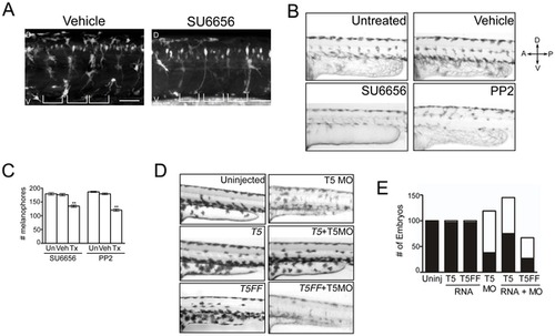
Neural crest derivatives require a Src-Tks5-dependent pathway in vivo. (A) Tg(sox10:RFP) embryos (8 hpf) were treated with either vehicle (DMSO) or SU6656 for 24 hours and imaged by confocal microscopy to detect neural crest cells. (D = dorsal, V = ventral). Brackets indicate the position of the somites. Scale bar represents 50 μm. (B–C) Embryos at 15 hpf were treated as indicated for 24 hours and analyzed for pigmentation defects. (B) Embryos where treatment was initiated at 15 hpf were examined for melanophore patterning in the trunk region above the yolk sac extension. (D = dorsal, V = ventral, A = anterior, P = posterior) (C) The total number of melanophores present in the dorsal and ventral pigment lines was counted for embryos within each group as described in Materials and Methods. Mean values (n = 3) and SEM were shown in graph. ** denotes p<0.01 for vehicle treated vs. SFK treated comparison. (D–E) Embryos were injected as indicated and qualitatively analyzed for defects described previously. Morpholino and RNA concentrations detailed in Materials and Methods. (E) Morphants were identified as described in Figure 1D and embryos within each group were quantified (white = morphants, black = normal). |